 Movie
Movie Controller


+ Open data

- Basic information
| Entry | Database: PDB / ID: 6xux | ||||||
|---|---|---|---|---|---|---|---|
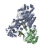
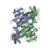
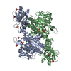
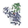
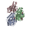
| Title | Crystal structure of Megabody Mb-Nb207-cYgjK_NO | ||||||
 Components Components | Nanobody,Glucosidase YgjK,Glucosidase YgjK,Nanobody | ||||||
 Keywords Keywords | STRUCTURAL PROTEIN / Megabody / Scaffold | ||||||
| Function / homology |  Function and homology information Function and homology informationglucosidase complex / alpha,alpha-trehalase activity / trehalose catabolic process / glucosidase activity / Hydrolases; Glycosylases; Glycosidases, i.e. enzymes that hydrolyse O- and S-glycosyl compounds / oligosaccharide catabolic process / DNA damage response / metal ion binding Similarity search - Function | ||||||
| Biological species |  | ||||||
| Method |  X-RAY DIFFRACTION / SYNCHROTRON / MOLECULAR REPLACEMENT / Resolution: 1.90000643078 Å X-RAY DIFFRACTION / SYNCHROTRON / MOLECULAR REPLACEMENT / Resolution: 1.90000643078 Å | ||||||
 Authors Authors | Steyaert, J. / Uchanski, T. / Fischer, B. | ||||||
| Funding support |  Belgium, 1items Belgium, 1items
| ||||||
 Citation Citation | Journal: Nat Methods / Year: 2021 Title: Megabodies expand the nanobody toolkit for protein structure determination by single-particle cryo-EM. Authors: Tomasz Uchański / Simonas Masiulis / Baptiste Fischer / Valentina Kalichuk / Uriel López-Sánchez / Eleftherios Zarkadas / Miriam Weckener / Andrija Sente / Philip Ward / Alexandre ...Authors: Tomasz Uchański / Simonas Masiulis / Baptiste Fischer / Valentina Kalichuk / Uriel López-Sánchez / Eleftherios Zarkadas / Miriam Weckener / Andrija Sente / Philip Ward / Alexandre Wohlkönig / Thomas Zögg / Han Remaut / James H Naismith / Hugues Nury / Wim Vranken / A Radu Aricescu / Els Pardon / Jan Steyaert /   Abstract: Nanobodies are popular and versatile tools for structural biology. They have a compact single immunoglobulin domain organization, bind target proteins with high affinities while reducing their ...Nanobodies are popular and versatile tools for structural biology. They have a compact single immunoglobulin domain organization, bind target proteins with high affinities while reducing their conformational heterogeneity and stabilize multi-protein complexes. Here we demonstrate that engineered nanobodies can also help overcome two major obstacles that limit the resolution of single-particle cryo-electron microscopy reconstructions: particle size and preferential orientation at the water-air interfaces. We have developed and characterized constructs, termed megabodies, by grafting nanobodies onto selected protein scaffolds to increase their molecular weight while retaining the full antigen-binding specificity and affinity. We show that the megabody design principles are applicable to different scaffold proteins and recognition domains of compatible geometries and are amenable for efficient selection from yeast display libraries. Moreover, we demonstrate that megabodies can be used to obtain three-dimensional reconstructions for membrane proteins that suffer from severe preferential orientation or are otherwise too small to allow accurate particle alignment. | ||||||
| History |
|
- Structure visualization
| Structure viewer | Molecule: MolmilJmol/JSmol |
|---|
- Downloads & links
-Download
| PDBx/mmCIF format | 6xux.cif.gz | 207.8 KB | Display | PDBx/mmCIF format |
|---|---|---|---|---|
| PDB format | pdb6xux.ent.gz | 158.8 KB | Display | PDB format |
| PDBx/mmJSON format | 6xux.json.gz | Tree view | PDBx/mmJSON format | |
| Others |  Other downloads Other downloads |
-Validation report
| Arichive directory | https://data.pdbj.org/pub/pdb/validation_reports/xu/6xuxftp://data.pdbj.org/pub/pdb/validation_reports/xu/6xux | HTTPS FTP |
|---|
-Related structure data
| Related structure data |  4542C 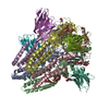 6qfaC  6xv8C  6xviC 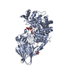 3w7sS S: Starting model for refinement C: citing same article ( |
|---|---|
| Similar structure data |


-Links
 PDBj
PDBj


- Assembly
| Deposited unit | 
| ||||||||||||
|---|---|---|---|---|---|---|---|---|---|---|---|---|---|
| 1 |
| ||||||||||||
| Unit cell |
|
-Components
| #1: Antibody | Mass: 101619.000 Da / Num. of mol.: 1 / Fragment: beta-strand A Source method: isolated from a genetically manipulated source Source: (gene. exp.) References: UniProt: P42592, Hydrolases; Glycosylases; Glycosidases, i.e. enzymes that hydrolyse O- and S-glycosyl compounds |
|---|---|
| #2: Chemical | ChemComp-CA /   Mass: 40.078 Da / Num. of mol.: 1 / Source method: obtained synthetically / Formula: Ca Mass: 40.078 Da / Num. of mol.: 1 / Source method: obtained synthetically / Formula: Ca |
| #3: Water | ChemComp-HOH /  Mass: 18.015 Da / Num. of mol.: 650 / Source method: isolated from a natural source / Formula: H2O Mass: 18.015 Da / Num. of mol.: 650 / Source method: isolated from a natural source / Formula: H2O |
| Has ligand of interest | N |
| Has protein modification | Y |
-Experimental details
-Experiment
| Experiment | Method: X-RAY DIFFRACTION / Number of used crystals: 1 |
|---|
- Sample preparation
| Crystal | Density Matthews: 1.96 Å3/Da / Density % sol: 37.2 % |
|---|---|
| Crystal grow | Temperature: 293 K / Method: vapor diffusion Details: 0.1 M Sodium citrate pH 7.5, 20% PEG 8000, 20% glycerol |
-Data collection
| Diffraction | Mean temperature: 100 K / Serial crystal experiment: N |
|---|---|
| Diffraction source | Source: SYNCHROTRON / Site: Diamond / Beamline: I03 / Wavelength: 0.9763 Å |
| Detector | Type: DECTRIS EIGER2 XE 16M / Detector: PIXEL / Date: May 20, 2019 |
| Radiation | Protocol: SINGLE WAVELENGTH / Monochromatic (M) / Laue (L): M / Scattering type: x-ray |
| Radiation wavelength | Wavelength: 0.9763 Å / Relative weight: 1 |
| Reflection | Resolution: 1.9→42.15 Å / Num. obs: 63885 / % possible obs: 100 % / Redundancy: 1.999 % / Biso Wilson estimate: 21.2241179387 Å2 / CC1/2: 0.998 / Rmerge(I) obs: 0.038 / Rrim(I) all: 0.054 / Net I/σ(I): 11.1 |
| Reflection shell | Resolution: 1.9→1.97 Å / Redundancy: 1.999 % / Rmerge(I) obs: 0.234 / Mean I/σ(I) obs: 3.4 / Num. unique obs: 6311 / CC1/2: 0.869 / Rrim(I) all: 0.331 / % possible all: 100 |
- Processing
| Software |
| |||||||||||||||||||||||||||||||||||||||||||||||||||||||||||||||||||||||||||||||||||||||||||||||||||||||||||||||||||||||||||||||||||||||||||||||||||||||||||||||||
|---|---|---|---|---|---|---|---|---|---|---|---|---|---|---|---|---|---|---|---|---|---|---|---|---|---|---|---|---|---|---|---|---|---|---|---|---|---|---|---|---|---|---|---|---|---|---|---|---|---|---|---|---|---|---|---|---|---|---|---|---|---|---|---|---|---|---|---|---|---|---|---|---|---|---|---|---|---|---|---|---|---|---|---|---|---|---|---|---|---|---|---|---|---|---|---|---|---|---|---|---|---|---|---|---|---|---|---|---|---|---|---|---|---|---|---|---|---|---|---|---|---|---|---|---|---|---|---|---|---|---|---|---|---|---|---|---|---|---|---|---|---|---|---|---|---|---|---|---|---|---|---|---|---|---|---|---|---|---|---|---|---|---|
| Refinement | Method to determine structure: MOLECULAR REPLACEMENT Starting model: 3W7S Resolution: 1.90000643078→42.1397385191 Å / SU ML: 0.194259667698 / Cross valid method: FREE R-VALUE / σ(F): 1.34351852722 / Phase error: 25.5416642491
| |||||||||||||||||||||||||||||||||||||||||||||||||||||||||||||||||||||||||||||||||||||||||||||||||||||||||||||||||||||||||||||||||||||||||||||||||||||||||||||||||
| Solvent computation | Shrinkage radii: 0.9 Å / VDW probe radii: 1.11 Å | |||||||||||||||||||||||||||||||||||||||||||||||||||||||||||||||||||||||||||||||||||||||||||||||||||||||||||||||||||||||||||||||||||||||||||||||||||||||||||||||||
| Displacement parameters | Biso mean: 26.5806085483 Å2 | |||||||||||||||||||||||||||||||||||||||||||||||||||||||||||||||||||||||||||||||||||||||||||||||||||||||||||||||||||||||||||||||||||||||||||||||||||||||||||||||||
| Refinement step | Cycle: LAST / Resolution: 1.90000643078→42.1397385191 Å
| |||||||||||||||||||||||||||||||||||||||||||||||||||||||||||||||||||||||||||||||||||||||||||||||||||||||||||||||||||||||||||||||||||||||||||||||||||||||||||||||||
| Refine LS restraints |
| |||||||||||||||||||||||||||||||||||||||||||||||||||||||||||||||||||||||||||||||||||||||||||||||||||||||||||||||||||||||||||||||||||||||||||||||||||||||||||||||||
| LS refinement shell |
|
