Journal: Nat Methods / Year: 2021 Title: Megabodies expand the nanobody toolkit for protein structure determination by single-particle cryo-EM. Authors: Tomasz Uchański / Simonas Masiulis / Baptiste Fischer / Valentina Kalichuk / Uriel López-Sánchez / Eleftherios Zarkadas / Miriam Weckener / Andrija Sente / Philip Ward / Alexandre ...Authors: Tomasz Uchański / Simonas Masiulis / Baptiste Fischer / Valentina Kalichuk / Uriel López-Sánchez / Eleftherios Zarkadas / Miriam Weckener / Andrija Sente / Philip Ward / Alexandre Wohlkönig / Thomas Zögg / Han Remaut / James H Naismith / Hugues Nury / Wim Vranken / A Radu Aricescu / Els Pardon / Jan Steyaert / Abstract: Nanobodies are popular and versatile tools for structural biology. They have a compact single immunoglobulin domain organization, bind target proteins with high affinities while reducing their ...Nanobodies are popular and versatile tools for structural biology. They have a compact single immunoglobulin domain organization, bind target proteins with high affinities while reducing their conformational heterogeneity and stabilize multi-protein complexes. Here we demonstrate that engineered nanobodies can also help overcome two major obstacles that limit the resolution of single-particle cryo-electron microscopy reconstructions: particle size and preferential orientation at the water-air interfaces. We have developed and characterized constructs, termed megabodies, by grafting nanobodies onto selected protein scaffolds to increase their molecular weight while retaining the full antigen-binding specificity and affinity. We show that the megabody design principles are applicable to different scaffold proteins and recognition domains of compatible geometries and are amenable for efficient selection from yeast display libraries. Moreover, we demonstrate that megabodies can be used to obtain three-dimensional reconstructions for membrane proteins that suffer from severe preferential orientation or are otherwise too small to allow accurate particle alignment.
Mass: 56165.656 Da / Num. of mol.: 4 Source method: isolated from a genetically manipulated source Source: (gene. exp.) Lama glama (llama), (gene. exp.) Helicobacter pylori (strain G27) (bacteria) Gene: Nb207, hopQ, HPG27_1120 / Strain: G27 / Production host: Escherichia coli (E. coli) / References: UniProt: B5Z8H1
Has protein modification
Y
Sequence details
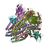
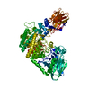
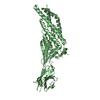
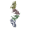
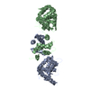
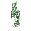
Megabody Mb-Nb207-c7HopQ_G10 is a chimeric protein with circular permutation of HopQ: Residues 1-12: ...Megabody Mb-Nb207-c7HopQ_G10 is a chimeric protein with circular permutation of HopQ: Residues 1-12: a part of a β-strand A of the Nanobody fold. Residue 13: one amino acid linker. Residues 14-232: C-terminal part of HopQ (residues 228-446, UniProtKB B5Z8H1). Residues 233-400: N-terminal part of HopQ (residues 53-220, UniProtKB B5Z8H1). Residue 401: one amino acid linker. Residues 402-511: a part of the Nanobody fold. Residues 512-517: the His6 tag. Residues 518-521: the EPEA tag.
-
Experimental details
-
Experiment
Experiment
Method: X-RAY DIFFRACTION / Number of used crystals: 1
-
Sample preparation
Crystal
Density Matthews: 2.35 Å3/Da / Density % sol: 47.7 %
Crystal grow
Temperature: 293 K / Method: vapor diffusion / pH: 7.5 / Details: 0.1 M MgCl2, 0.1 M HEPES pH 7.5, 19% PEG 4000
-
Data collection
Diffraction
Mean temperature: 100 K / Serial crystal experiment: N
In the structure databanks used in Yorodumi, some data are registered as the other names, "COVID-19 virus" and "2019-nCoV". Here are the details of the virus and the list of structure data.
Jan 31, 2019. EMDB accession codes are about to change! (news from PDBe EMDB page)
EMDB accession codes are about to change! (news from PDBe EMDB page)
The allocation of 4 digits for EMDB accession codes will soon come to an end. Whilst these codes will remain in use, new EMDB accession codes will include an additional digit and will expand incrementally as the available range of codes is exhausted. The current 4-digit format prefixed with “EMD-” (i.e. EMD-XXXX) will advance to a 5-digit format (i.e. EMD-XXXXX), and so on. It is currently estimated that the 4-digit codes will be depleted around Spring 2019, at which point the 5-digit format will come into force.
The EM Navigator/Yorodumi systems omit the EMD- prefix.
Related info.:Q: What is EMD? / ID/Accession-code notation in Yorodumi/EM Navigator
Yorodumi is a browser for structure data from EMDB, PDB, SASBDB, etc.
This page is also the successor to EM Navigator detail page, and also detail information page/front-end page for Omokage search.
The word "yorodu" (or yorozu) is an old Japanese word meaning "ten thousand". "mi" (miru) is to see.
Related info.:EMDB / PDB / SASBDB / Comparison of 3 databanks / Yorodumi Search / Aug 31, 2016. New EM Navigator & Yorodumi / Yorodumi Papers / Jmol/JSmol / Function and homology information / Changes in new EM Navigator and Yorodumi
 Movie
Movie Controller


 Open data
 Basic information
 Components
 Keywords
 Function and homology information

 Helicobacter pylori (bacteria)
 X-RAY DIFFRACTION /
  Authors
 Belgium, 1items
  Citation

 Structure visualization
 Downloads & links
 Other downloads


 PDBj
PDBj

 Assembly
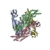
 Sample preparation
 Processing