 Movie
Movie Controller

[English] 日本語
 Yorodumi
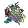
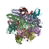
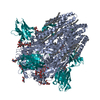
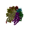
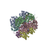
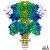
- PDB-6qfa: CryoEM structure of a beta3K279T GABA(A)R homomer in complex with... -
+ Open data

- Basic information
| Entry | Database: PDB / ID: 6qfa | ||||||||||||||||||
|---|---|---|---|---|---|---|---|---|---|---|---|---|---|---|---|---|---|---|---|
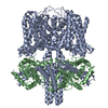
| Title | CryoEM structure of a beta3K279T GABA(A)R homomer in complex with histamine and megabody Mb25 | ||||||||||||||||||
 Components Components |
| ||||||||||||||||||
 Keywords Keywords | MEMBRANE PROTEIN / Megabody / protein engineering / cryo-EM | ||||||||||||||||||
| Function / homology |  Function and homology information Function and homology informationcircadian sleep/wake cycle, REM sleep / reproductive behavior / hard palate development / cellular response to histamine / GABA receptor activation / inner ear receptor cell development / GABA-gated chloride ion channel activity / GABA-A receptor complex / inhibitory synapse assembly / innervation ...circadian sleep/wake cycle, REM sleep / reproductive behavior / hard palate development / cellular response to histamine / GABA receptor activation / inner ear receptor cell development / GABA-gated chloride ion channel activity / GABA-A receptor complex / inhibitory synapse assembly / innervation / GABA-A receptor activity / response to anesthetic / postsynaptic specialization membrane / gamma-aminobutyric acid signaling pathway / synaptic transmission, GABAergic / motor behavior / inhibitory postsynaptic potential / cellular response to zinc ion / roof of mouth development / exploration behavior / Signaling by ERBB4 / cochlea development / social behavior / chloride channel complex / cerebellum development / cytoplasmic vesicle membrane / chloride transmembrane transport / learning / transmitter-gated monoatomic ion channel activity involved in regulation of postsynaptic membrane potential / GABA-ergic synapse / memory / dendritic spine / postsynaptic membrane / response to xenobiotic stimulus / cell surface / signal transduction / identical protein binding / plasma membrane Similarity search - Function | ||||||||||||||||||
| Biological species |  Homo sapiens (human) Homo sapiens (human)  Helicobacter pylori (bacteria) Helicobacter pylori (bacteria) | ||||||||||||||||||
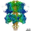
| Method | ELECTRON MICROSCOPY / single particle reconstruction / cryo EM / Resolution: 2.49 Å | ||||||||||||||||||
 Authors Authors | Uchanski, T. / Masiulis, S. / Fischer, B. / Kalichuk, V. / Wohlkoening, A. / Zoegg, T. / Remaut, H. / Vranken, W. / Aricescu, A.R. / Pardon, E. / Steyaert, J. | ||||||||||||||||||
| Funding support |  United Kingdom, United Kingdom,  Belgium, 5items Belgium, 5items
| ||||||||||||||||||
 Citation Citation | Journal: Nat Methods / Year: 2021 Title: Megabodies expand the nanobody toolkit for protein structure determination by single-particle cryo-EM. Authors: Tomasz Uchański / Simonas Masiulis / Baptiste Fischer / Valentina Kalichuk / Uriel López-Sánchez / Eleftherios Zarkadas / Miriam Weckener / Andrija Sente / Philip Ward / Alexandre ...Authors: Tomasz Uchański / Simonas Masiulis / Baptiste Fischer / Valentina Kalichuk / Uriel López-Sánchez / Eleftherios Zarkadas / Miriam Weckener / Andrija Sente / Philip Ward / Alexandre Wohlkönig / Thomas Zögg / Han Remaut / James H Naismith / Hugues Nury / Wim Vranken / A Radu Aricescu / Els Pardon / Jan Steyaert /  Abstract: Nanobodies are popular and versatile tools for structural biology. They have a compact single immunoglobulin domain organization, bind target proteins with high affinities while reducing their ...Nanobodies are popular and versatile tools for structural biology. They have a compact single immunoglobulin domain organization, bind target proteins with high affinities while reducing their conformational heterogeneity and stabilize multi-protein complexes. Here we demonstrate that engineered nanobodies can also help overcome two major obstacles that limit the resolution of single-particle cryo-electron microscopy reconstructions: particle size and preferential orientation at the water-air interfaces. We have developed and characterized constructs, termed megabodies, by grafting nanobodies onto selected protein scaffolds to increase their molecular weight while retaining the full antigen-binding specificity and affinity. We show that the megabody design principles are applicable to different scaffold proteins and recognition domains of compatible geometries and are amenable for efficient selection from yeast display libraries. Moreover, we demonstrate that megabodies can be used to obtain three-dimensional reconstructions for membrane proteins that suffer from severe preferential orientation or are otherwise too small to allow accurate particle alignment. | ||||||||||||||||||
| History |
|
- Structure visualization
| Movie |
Movie viewer |
|---|---|
| Structure viewer | Molecule: MolmilJmol/JSmol |
- Downloads & links
-Download
| PDBx/mmCIF format | 6qfa.cif.gz | 439.4 KB | Display | PDBx/mmCIF format |
|---|---|---|---|---|
| PDB format | pdb6qfa.ent.gz | 348.2 KB | Display | PDB format |
| PDBx/mmJSON format | 6qfa.json.gz | Tree view | PDBx/mmJSON format | |
| Others |  Other downloads Other downloads |
-Validation report
| Arichive directory | https://data.pdbj.org/pub/pdb/validation_reports/qf/6qfaftp://data.pdbj.org/pub/pdb/validation_reports/qf/6qfa | HTTPS FTP |
|---|
-Related structure data
| Related structure data |  4542MC 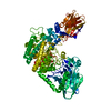 6xuxC  6xv8C  6xviC M: map data used to model this data C: citing same article ( |
|---|---|
| Similar structure data |

-Links
 PDBj
PDBj


- Assembly
| Deposited unit | 
|
|---|---|
| 1 |
|
-Components
| #1: Protein | Mass: 39503.555 Da / Num. of mol.: 5 / Mutation: K279T Source method: isolated from a genetically manipulated source Source: (gene. exp.) Homo sapiens (human) / Gene: GABRB3 / Cell line (production host): HEK293S GnTI- / Production host: Homo sapiens (human) / References: UniProt: P28472#2: Antibody | Mass: 56300.629 Da / Num. of mol.: 5 Source method: isolated from a genetically manipulated source Source: (gene. exp.) Helicobacter pylori (strain G27) (bacteria), (gene. exp.) Strain: G27 / Gene: hopQ, HPG27_1120 / Production host: #3: Polysaccharide | alpha-D-mannopyranose-(1-3)-[alpha-D-mannopyranose-(1-6)]beta-D-mannopyranose-(1-4)-2-acetamido-2- ...alpha-D-mannopyranose-(1-3)-[alpha-D-mannopyranose-(1-6)]beta-D-mannopyranose-(1-4)-2-acetamido-2-deoxy-beta-D-glucopyranose-(1-4)-2-acetamido-2-deoxy-beta-D-glucopyranose Source method: isolated from a genetically manipulated source #4: Sugar | ChemComp-NAG /   Type: D-saccharide, beta linking / Mass: 221.208 Da / Num. of mol.: 5 Type: D-saccharide, beta linking / Mass: 221.208 Da / Num. of mol.: 5Source method: isolated from a genetically manipulated source Formula: C8H15NO6 #5: Chemical | ChemComp-HSM / 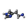  Mass: 111.145 Da / Num. of mol.: 5 Mass: 111.145 Da / Num. of mol.: 5Source method: isolated from a genetically manipulated source Formula: C5H9N3 / Feature type: SUBJECT OF INVESTIGATION Has protein modification | Y | |
|---|
-Experimental details
-Experiment
| Experiment | Method: ELECTRON MICROSCOPY |
|---|---|
| EM experiment | Aggregation state: PARTICLE / 3D reconstruction method: single particle reconstruction |
- Sample preparation
| Component |
| ||||||||||||||||||||||||
|---|---|---|---|---|---|---|---|---|---|---|---|---|---|---|---|---|---|---|---|---|---|---|---|---|---|
| Molecular weight | Value: 0.5 MDa / Experimental value: NO | ||||||||||||||||||||||||
| Source (natural) |
| ||||||||||||||||||||||||
| Source (recombinant) |
| ||||||||||||||||||||||||
| Buffer solution | pH: 7.6 | ||||||||||||||||||||||||
| Specimen | Conc.: 0.1 mg/ml / Embedding applied: NO / Shadowing applied: NO / Staining applied: NO / Vitrification applied: YES / Details: Monodisperse sample | ||||||||||||||||||||||||
| Vitrification | Instrument: FEI VITROBOT MARK IV / Cryogen name: ETHANE |
- Electron microscopy imaging
| Experimental equipment |  Model: Titan Krios / Image courtesy: FEI Company |
|---|---|
| Microscopy | Model: FEI TITAN KRIOS |
| Electron gun | Electron source:  FIELD EMISSION GUN / Accelerating voltage: 300 kV / Illumination mode: FLOOD BEAM FIELD EMISSION GUN / Accelerating voltage: 300 kV / Illumination mode: FLOOD BEAM |
| Electron lens | Mode: BRIGHT FIELD / Nominal magnification: 75000 X / Nominal defocus max: 700 nm / Nominal defocus min: 500 nm / Cs: 2.7 mm |
| Specimen holder | Cryogen: NITROGEN / Specimen holder model: FEI TITAN KRIOS AUTOGRID HOLDER |
| Image recording | Electron dose: 30 e/Å2 / Detector mode: COUNTING / Film or detector model: FEI FALCON III (4k x 4k) |
| EM imaging optics | Phase plate: VOLTA PHASE PLATE |
- Processing
| EM software |
| |||||||||||||||||||||||||||||||||||||||||||||
|---|---|---|---|---|---|---|---|---|---|---|---|---|---|---|---|---|---|---|---|---|---|---|---|---|---|---|---|---|---|---|---|---|---|---|---|---|---|---|---|---|---|---|---|---|---|---|
| CTF correction | Type: PHASE FLIPPING AND AMPLITUDE CORRECTION | |||||||||||||||||||||||||||||||||||||||||||||
| Particle selection | Num. of particles selected: 315183 | |||||||||||||||||||||||||||||||||||||||||||||
| Symmetry | Point symmetry: C5 (5 fold cyclic) | |||||||||||||||||||||||||||||||||||||||||||||
| 3D reconstruction | Resolution: 2.49 Å / Resolution method: FSC 0.143 CUT-OFF / Num. of particles: 171761 / Algorithm: BACK PROJECTION / Symmetry type: POINT | |||||||||||||||||||||||||||||||||||||||||||||
| Atomic model building | Protocol: FLEXIBLE FIT |
