 Movie
Movie Controller

[English] 日本語
 Yorodumi
- PDB-6xpq: Human antibody D1 H1-17/H3-14 in complex with the influenza hemag... -
+ Open data

- Basic information
| Entry | Database: PDB / ID: 6xpq | ||||||||||||
|---|---|---|---|---|---|---|---|---|---|---|---|---|---|
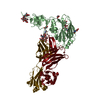
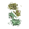
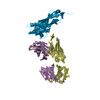
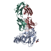
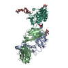
| Title | Human antibody D1 H1-17/H3-14 in complex with the influenza hemagglutinin head domain of A/Texas/50/2012(H3N2) | ||||||||||||
 Components Components |
| ||||||||||||
 Keywords Keywords | VIRAL PROTEIN/IMMUNE SYSTEM / antibody antigen complex / influenza / hemagglutinin / VIRAL PROTEIN-IMMUNE SYSTEM complex | ||||||||||||
| Function / homology |  Function and homology information Function and homology informationviral budding from plasma membrane / clathrin-dependent endocytosis of virus by host cell / host cell surface receptor binding / fusion of virus membrane with host plasma membrane / fusion of virus membrane with host endosome membrane / viral envelope / virion attachment to host cell / host cell plasma membrane / virion membrane / membrane Similarity search - Function | ||||||||||||
| Biological species |   Influenza A virus Influenza A virus Homo sapiens (human) Homo sapiens (human) | ||||||||||||
| Method |  X-RAY DIFFRACTION / SYNCHROTRON / MOLECULAR REPLACEMENT / Resolution: 4.2 Å X-RAY DIFFRACTION / SYNCHROTRON / MOLECULAR REPLACEMENT / Resolution: 4.2 Å | ||||||||||||
 Authors Authors | McCarthy, K.R. / Harrison, S.C. | ||||||||||||
| Funding support |  United States, 3items United States, 3items
| ||||||||||||
 Citation Citation | Journal: Mbio / Year: 2021 Title: A Prevalent Focused Human Antibody Response to the Influenza Virus Hemagglutinin Head Interface. Authors: McCarthy, K.R. / Lee, J. / Watanabe, A. / Kuraoka, M. / Robinson-McCarthy, L.R. / Georgiou, G. / Kelsoe, G. / Harrison, S.C. | ||||||||||||
| History |
|
- Structure visualization
| Structure viewer | Molecule: MolmilJmol/JSmol |
|---|
- Downloads & links
-Download
| PDBx/mmCIF format | 6xpq.cif.gz | 156.5 KB | Display | PDBx/mmCIF format |
|---|---|---|---|---|
| PDB format | pdb6xpq.ent.gz | 119.7 KB | Display | PDB format |
| PDBx/mmJSON format | 6xpq.json.gz | Tree view | PDBx/mmJSON format | |
| Others |  Other downloads Other downloads |
-Validation report
| Arichive directory | https://data.pdbj.org/pub/pdb/validation_reports/xp/6xpqftp://data.pdbj.org/pub/pdb/validation_reports/xp/6xpq | HTTPS FTP |
|---|
-Related structure data
| Related structure data |  6xpoC  6xprC  6xpxC  6xpyC  6xpzC  6xq0C  6xq2C  6xq4C  5w08S  6e4xS S: Starting model for refinement C: citing same article ( |
|---|---|
| Similar structure data |


-Links
 PDBj
PDBj


- Assembly
| Deposited unit | 
| ||||||||
|---|---|---|---|---|---|---|---|---|---|
| 1 |
| ||||||||
| Unit cell |
|
-Components
| #1: Protein | Mass: 32618.846 Da / Num. of mol.: 1 / Fragment: head domain (UNP residues 53-335) Source method: isolated from a genetically manipulated source Source: (gene. exp.) Influenza A virus (A/Texas/50/2012(H3N2))Strain: A/Texas/50/2012(H3N2) / Gene: HA, L998_47834gpHA / Cell line (production host): BTI-Tn-5B1-4 / Production host:  Trichoplusia ni (cabbage looper) / References: UniProt: R4L1D1 Trichoplusia ni (cabbage looper) / References: UniProt: R4L1D1 | ||||
|---|---|---|---|---|---|
| #2: Antibody | Mass: 23168.607 Da / Num. of mol.: 1 Source method: isolated from a genetically manipulated source Source: (gene. exp.) Homo sapiens (human) / Cell line (production host): 293F / Production host: Homo sapiens (human) | ||||
| #3: Antibody | Mass: 26629.750 Da / Num. of mol.: 1 Source method: isolated from a genetically manipulated source Source: (gene. exp.) Homo sapiens (human) / Cell line (production host): 293F / Production host: Homo sapiens (human) | ||||
| #4: Polysaccharide | Source method: isolated from a genetically manipulated source Has ligand of interest | N | Has protein modification | Y | |
-Experimental details
-Experiment
| Experiment | Method: X-RAY DIFFRACTION / Number of used crystals: 1 |
|---|
- Sample preparation
| Crystal | Density Matthews: 5.76 Å3/Da / Density % sol: 78.66 % |
|---|---|
| Crystal grow | Temperature: 291.15 K / Method: vapor diffusion, hanging drop / Details: 0.5 M lithium chloride, 1.0 M sodium citrate |
-Data collection
| Diffraction | Mean temperature: 80 K / Serial crystal experiment: N |
|---|---|
| Diffraction source | Source: SYNCHROTRON / Site: APS / Beamline: 24-ID-E / Wavelength: 0.97918 Å |
| Detector | Type: DECTRIS EIGER X 16M / Detector: PIXEL / Date: Mar 26, 2019 |
| Radiation | Monochromator: single crystal Si(220) side bounce / Protocol: SINGLE WAVELENGTH / Monochromatic (M) / Laue (L): M / Scattering type: x-ray |
| Radiation wavelength | Wavelength: 0.97918 Å / Relative weight: 1 |
| Reflection | Resolution: 4.2→49.105 Å / Num. obs: 13796 / % possible obs: 96.58 % / Redundancy: 3.7 % / CC1/2: 0.997 / CC star: 0.999 / Rmerge(I) obs: 0.1066 / Rpim(I) all: 0.06226 / Rrim(I) all: 0.1241 / Net I/σ(I): 7.62 |
| Reflection shell | Resolution: 4.2→4.35 Å / Redundancy: 3.8 % / Rmerge(I) obs: 0.9088 / Mean I/σ(I) obs: 1.19 / Num. unique obs: 1386 / CC1/2: 0.639 / CC star: 0.883 / Rpim(I) all: 0.5109 / Rrim(I) all: 1.047 / % possible all: 98.16 |
- Processing
| Software |
| ||||||||||||||||||||||||||||||||||||
|---|---|---|---|---|---|---|---|---|---|---|---|---|---|---|---|---|---|---|---|---|---|---|---|---|---|---|---|---|---|---|---|---|---|---|---|---|---|
| Refinement | Method to determine structure: MOLECULAR REPLACEMENT Starting model: PDB entries 5W08 & 6E4X Resolution: 4.2→49.105 Å / SU ML: 0.69 / Cross valid method: THROUGHOUT / σ(F): 1.34 / Phase error: 36.75 / Stereochemistry target values: ML
| ||||||||||||||||||||||||||||||||||||
| Solvent computation | Shrinkage radii: 0.9 Å / VDW probe radii: 1.11 Å / Solvent model: FLAT BULK SOLVENT MODEL | ||||||||||||||||||||||||||||||||||||
| Displacement parameters | Biso max: 384.83 Å2 / Biso mean: 236.6196 Å2 / Biso min: 133.66 Å2 | ||||||||||||||||||||||||||||||||||||
| Refinement step | Cycle: final / Resolution: 4.2→49.105 Å
| ||||||||||||||||||||||||||||||||||||
| LS refinement shell | Refine-ID: X-RAY DIFFRACTION / Rfactor Rfree error: 0
|
