 Movie
Movie Controller

[English] 日本語
 Yorodumi
- PDB-6wg7: Coordinates of NanR dimer fitted in Hexameric NanR-DNA hetero-com... -
+ Open data

- Basic information
| Entry | Database: PDB / ID: 6wg7 | ||||||
|---|---|---|---|---|---|---|---|
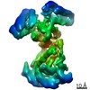
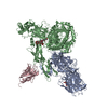
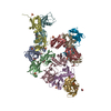
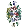
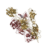
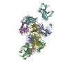
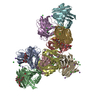
| Title | Coordinates of NanR dimer fitted in Hexameric NanR-DNA hetero-complex cryo-EM map | ||||||
 Components Components |
| ||||||
 Keywords Keywords | GENE REGULATION / NanR dimer-DNA hetero-complex / transcriptional regulator / GntR superfamily / sialic acid / Neu5Ac / cooperativity / Gene regulation. | ||||||
| Function / homology |  Function and homology information Function and homology informationDNA-binding transcription factor activity / negative regulation of DNA-templated transcription / DNA binding Similarity search - Function | ||||||
| Biological species |  synthetic construct (others) | ||||||
| Method | ELECTRON MICROSCOPY / single particle reconstruction / cryo EM / Resolution: 8.3 Å | ||||||
 Authors Authors | Hariprasad, V. / Horne, C. / Santosh, P. / Amy, H. / Emre, B. / Rachel, N. / Michael, G. / Georg, R. / Borries, D. / Renwick, D. | ||||||
| Funding support |  New Zealand, 1items New Zealand, 1items
| ||||||
 Citation Citation | Journal: Nat Commun / Year: 2021 Title: Mechanism of NanR gene repression and allosteric induction of bacterial sialic acid metabolism. Authors: Christopher R Horne / Hariprasad Venugopal / Santosh Panjikar / David M Wood / Amy Henrickson / Emre Brookes / Rachel A North / James M Murphy / Rosmarie Friemann / Michael D W Griffin / ...Authors: Christopher R Horne / Hariprasad Venugopal / Santosh Panjikar / David M Wood / Amy Henrickson / Emre Brookes / Rachel A North / James M Murphy / Rosmarie Friemann / Michael D W Griffin / Georg Ramm / Borries Demeler / Renwick C J Dobson /     Abstract: Bacteria respond to environmental changes by inducing transcription of some genes and repressing others. Sialic acids, which coat human cell surfaces, are a nutrient source for pathogenic and ...Bacteria respond to environmental changes by inducing transcription of some genes and repressing others. Sialic acids, which coat human cell surfaces, are a nutrient source for pathogenic and commensal bacteria. The Escherichia coli GntR-type transcriptional repressor, NanR, regulates sialic acid metabolism, but the mechanism is unclear. Here, we demonstrate that three NanR dimers bind a (GGTATA)-repeat operator cooperatively and with high affinity. Single-particle cryo-electron microscopy structures reveal the DNA-binding domain is reorganized to engage DNA, while three dimers assemble in close proximity across the (GGTATA)-repeat operator. Such an interaction allows cooperative protein-protein interactions between NanR dimers via their N-terminal extensions. The effector, N-acetylneuraminate, binds NanR and attenuates the NanR-DNA interaction. The crystal structure of NanR in complex with N-acetylneuraminate reveals a domain rearrangement upon N-acetylneuraminate binding to lock NanR in a conformation that weakens DNA binding. Our data provide a molecular basis for the regulation of bacterial sialic acid metabolism. | ||||||
| History |
|
- Structure visualization
| Movie |
Movie viewer |
|---|---|
| Structure viewer | Molecule: MolmilJmol/JSmol |
- Downloads & links
-Download
| PDBx/mmCIF format | 6wg7.cif.gz | 278.6 KB | Display | PDBx/mmCIF format |
|---|---|---|---|---|
| PDB format | pdb6wg7.ent.gz | 218.4 KB | Display | PDB format |
| PDBx/mmJSON format | 6wg7.json.gz | Tree view | PDBx/mmJSON format | |
| Others |  Other downloads Other downloads |
-Validation report
| Arichive directory | https://data.pdbj.org/pub/pdb/validation_reports/wg/6wg7ftp://data.pdbj.org/pub/pdb/validation_reports/wg/6wg7 | HTTPS FTP |
|---|
-Related structure data
| Related structure data |  21661MC 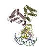 6wfqC C: citing same article ( M: map data used to model this data |
|---|---|
| Similar structure data |


-Links
 PDBj
PDBj


- Assembly
| Deposited unit | 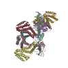
|
|---|---|
| 1 |
|
-Components
| #1: DNA chain | Mass: 10856.016 Da / Num. of mol.: 1 / Source method: obtained synthetically / Source: (synth.) synthetic construct (others) |
|---|---|
| #2: DNA chain | Mass: 10673.923 Da / Num. of mol.: 1 / Source method: obtained synthetically / Source: (synth.) synthetic construct (others) |
| #3: Protein | Mass: 29566.354 Da / Num. of mol.: 6 Source method: isolated from a genetically manipulated source Source: (gene. exp.) Production host: References: UniProt: J7QHT8 |
-Experimental details
-Experiment
| Experiment | Method: ELECTRON MICROSCOPY |
|---|---|
| EM experiment | Aggregation state: PARTICLE / 3D reconstruction method: single particle reconstruction |
- Sample preparation
| Component | Name: Hexameric NanR-DNA hetero-complex / Type: COMPLEX / Details: Hexameric NanR-DNA hetero-complex / Entity ID: all / Source: RECOMBINANT |
|---|---|
| Molecular weight | Value: 0.1985 MDa / Experimental value: YES |
| Source (natural) | Organism: |
| Source (recombinant) | Organism: |
| Buffer solution | pH: 8 / Details: 20mM Tris-HCL,50mM NaCl, pH 8.0 |
| Specimen | Conc.: 0.5 mg/ml / Embedding applied: NO / Shadowing applied: NO / Staining applied: NO / Vitrification applied: YES |
| Specimen support | Grid material: GOLD / Grid mesh size: 300 divisions/in. / Grid type: UltrAuFoil |
| Vitrification | Instrument: FEI VITROBOT MARK III / Cryogen name: ETHANE / Humidity: 100 % / Chamber temperature: 277.15 K / Details: Blot force: -3 Blot time: 3 sec Drain time: 0 |
- Electron microscopy imaging
| Microscopy | Model: TFS TALOS |
|---|---|
| Electron gun | Electron source:  FIELD EMISSION GUN / Accelerating voltage: 200 kV / Illumination mode: FLOOD BEAM FIELD EMISSION GUN / Accelerating voltage: 200 kV / Illumination mode: FLOOD BEAM |
| Electron lens | Mode: BRIGHT FIELD / Cs: 2.7 mm / C2 aperture diameter: 50 µm / Alignment procedure: COMA FREE |
| Specimen holder | Cryogen: NITROGEN / Specimen holder model: FEI TITAN KRIOS AUTOGRID HOLDER |
| Image recording | Average exposure time: 51.54 sec. / Electron dose: 45 e/Å2 / Detector mode: COUNTING / Film or detector model: FEI FALCON III (4k x 4k) / Num. of grids imaged: 1 / Num. of real images: 2287 |
- Processing
| EM software |
| ||||||||||||||||||||||||
|---|---|---|---|---|---|---|---|---|---|---|---|---|---|---|---|---|---|---|---|---|---|---|---|---|---|
| CTF correction | Type: PHASE FLIPPING AND AMPLITUDE CORRECTION | ||||||||||||||||||||||||
| Symmetry | Point symmetry: C1 (asymmetric) | ||||||||||||||||||||||||
| 3D reconstruction | Resolution: 8.3 Å / Resolution method: FSC 0.143 CUT-OFF / Num. of particles: 48931 / Symmetry type: POINT | ||||||||||||||||||||||||
| Atomic model building | Protocol: RIGID BODY FIT | ||||||||||||||||||||||||
| Atomic model building | 3D fitting-ID: 1 / Accession code: 6WFQ / Initial refinement model-ID: 1 / PDB-ID: 6WFQ / Source name: PDB / Type: experimental model
| ||||||||||||||||||||||||
| Refinement | Stereochemistry target values: GeoStd + Monomer Library + CDL v1.2 | ||||||||||||||||||||||||
| Displacement parameters | Biso mean: 247.92 Å2 | ||||||||||||||||||||||||
| Refine LS restraints |
|
