 Movie
Movie Controller

[English] 日本語
 Yorodumi
- PDB-6w00: Crystal structure of Fab239 in complex with NPNA2 peptide from ci... -
+ Open data

- Basic information
| Entry | Database: PDB / ID: 6w00 | ||||||
|---|---|---|---|---|---|---|---|
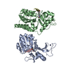
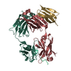
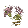
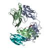
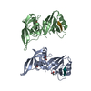
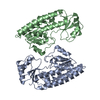
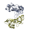
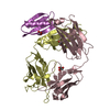
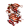
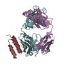
| Title | Crystal structure of Fab239 in complex with NPNA2 peptide from circumsporozoite protein | ||||||
 Components Components |
| ||||||
 Keywords Keywords | IMMUNE SYSTEM / Malaria / Sporozoite / Circumsporozoite protein / Antibody | ||||||
| Function / homology |  Function and homology information Function and homology informationentry into host cell by a symbiont-containing vacuole / IgG binding / side of membrane / cell surface / plasma membrane / cytoplasm Similarity search - Function | ||||||
| Biological species |  Streptococcus sp. group G (bacteria) Streptococcus sp. group G (bacteria) Homo sapiens (human) Homo sapiens (human) | ||||||
| Method |  X-RAY DIFFRACTION / SYNCHROTRON / MOLECULAR REPLACEMENT / Resolution: 1.853 Å X-RAY DIFFRACTION / SYNCHROTRON / MOLECULAR REPLACEMENT / Resolution: 1.853 Å | ||||||
 Authors Authors | Pholcharee, T. / Oyen, D. / Wilson, I.A. | ||||||
| Funding support |  United States, 1items United States, 1items
| ||||||
 Citation Citation | Journal: Nat Commun / Year: 2021 Title: Structural and biophysical correlation of anti-NANP antibodies with in vivo protection against P. falciparum. Authors: Pholcharee, T. / Oyen, D. / Flores-Garcia, Y. / Gonzalez-Paez, G. / Han, Z. / Williams, K.L. / Volkmuth, W. / Emerling, D. / Locke, E. / Richter King, C. / Zavala, F. / Wilson, I.A. #1: Journal: Biorxiv / Year: 2020Title: Structural and biophysical correlation of anti-NANP antibodies with in vivo protection against P. falciparum Authors: Pholcharee, T. / Oyen, D. / Flores-Garcia, Y. / Gonzalez-Paez, G. / Han, Z. / Williams, K.L. / Volkmuth, W. / Emerling, D. / Locke, E. / King, C.R. / Zavala, F. / Wilson, I.A. | ||||||
| History |
|
- Structure visualization
| Structure viewer | Molecule: MolmilJmol/JSmol |
|---|
- Downloads & links
-Download
| PDBx/mmCIF format | 6w00.cif.gz | 195.8 KB | Display | PDBx/mmCIF format |
|---|---|---|---|---|
| PDB format | pdb6w00.ent.gz | 155.9 KB | Display | PDB format |
| PDBx/mmJSON format | 6w00.json.gz | Tree view | PDBx/mmJSON format | |
| Others |  Other downloads Other downloads |
-Validation report
| Arichive directory | https://data.pdbj.org/pub/pdb/validation_reports/w0/6w00ftp://data.pdbj.org/pub/pdb/validation_reports/w0/6w00 | HTTPS FTP |
|---|
-Related structure data
| Related structure data |  6w05C  6wfwC  6wfxC  6wfyC  6wfzC  6wg0C  6wg1C  6wg2C C: citing same article ( |
|---|---|
| Similar structure data |
-Links
 PDBj
PDBj

- Assembly
| Deposited unit | 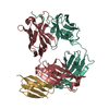
| ||||||||
|---|---|---|---|---|---|---|---|---|---|
| 1 |
| ||||||||
| Unit cell |
|
-Components
| #1: Antibody | Mass: 6327.939 Da / Num. of mol.: 1 / Fragment: domain III (UNP residues 438-497) Source method: isolated from a genetically manipulated source Source: (gene. exp.) Streptococcus sp. group G (bacteria) / Gene: spg / Production host: |
|---|---|
| #2: Antibody | Mass: 23614.164 Da / Num. of mol.: 1 Source method: isolated from a genetically manipulated source Source: (gene. exp.) Homo sapiens (human) / Production host:   Cricetulus griseus (Chinese hamster) Cricetulus griseus (Chinese hamster) |
| #3: Antibody | Mass: 24197.186 Da / Num. of mol.: 1 Source method: isolated from a genetically manipulated source Source: (gene. exp.) Homo sapiens (human) / Production host: Cricetulus griseus (Chinese hamster) |
| #4: Protein/peptide | Mass: 836.849 Da / Num. of mol.: 1 / Source method: obtained synthetically Source: (synth.) References: UniProt: P02893*PLUS |
| #5: Water | ChemComp-HOH /  Mass: 18.015 Da / Num. of mol.: 349 / Source method: isolated from a natural source / Formula: H2O Mass: 18.015 Da / Num. of mol.: 349 / Source method: isolated from a natural source / Formula: H2O |
| Has ligand of interest | N |
| Has protein modification | Y |
-Experimental details
-Experiment
| Experiment | Method: X-RAY DIFFRACTION / Number of used crystals: 1 |
|---|
- Sample preparation
| Crystal | Density Matthews: 2.77 Å3/Da / Density % sol: 55.65 % |
|---|---|
| Crystal grow | Temperature: 293.15 K / Method: vapor diffusion, sitting drop / Details: 0.2 M sodium chloride, 20% w/v PEG3350 |
-Data collection
| Diffraction | Mean temperature: 100 K / Serial crystal experiment: N |
|---|---|
| Diffraction source | Source: SYNCHROTRON / Site: APS / Beamline: 23-ID-D / Wavelength: 1.03321 Å |
| Detector | Type: DECTRIS PILATUS3 6M / Detector: PIXEL / Date: Oct 26, 2018 |
| Radiation | Monochromator: double crystal cryo-cooled Si(111) / Protocol: SINGLE WAVELENGTH / Monochromatic (M) / Laue (L): M / Scattering type: x-ray |
| Radiation wavelength | Wavelength: 1.03321 Å / Relative weight: 1 |
| Reflection | Resolution: 1.85→50 Å / Num. obs: 52998 / % possible obs: 100 % / Redundancy: 13.3 % / CC1/2: 0.951 / Net I/σ(I): 18.6 |
| Reflection shell | Resolution: 1.85→1.88 Å / Mean I/σ(I) obs: 1.9 / Num. unique obs: 2623 / CC1/2: 0.725 / Rpim(I) all: 0.303 / Rsym value: 0.933 |
- Processing
| Software |
| |||||||||||||||||||||||||||||||||||||||||||||||||||||||||||||||||||||||||||||||||||||||||||||||||||||||||||||||||||||||||||||||||||||
|---|---|---|---|---|---|---|---|---|---|---|---|---|---|---|---|---|---|---|---|---|---|---|---|---|---|---|---|---|---|---|---|---|---|---|---|---|---|---|---|---|---|---|---|---|---|---|---|---|---|---|---|---|---|---|---|---|---|---|---|---|---|---|---|---|---|---|---|---|---|---|---|---|---|---|---|---|---|---|---|---|---|---|---|---|---|---|---|---|---|---|---|---|---|---|---|---|---|---|---|---|---|---|---|---|---|---|---|---|---|---|---|---|---|---|---|---|---|---|---|---|---|---|---|---|---|---|---|---|---|---|---|---|---|---|
| Refinement | Method to determine structure: MOLECULAR REPLACEMENT Starting model: Homology model Resolution: 1.853→48.946 Å / SU ML: 0.18 / Cross valid method: FREE R-VALUE / σ(F): 1.36 / Phase error: 18.76 / Stereochemistry target values: ML
| |||||||||||||||||||||||||||||||||||||||||||||||||||||||||||||||||||||||||||||||||||||||||||||||||||||||||||||||||||||||||||||||||||||
| Solvent computation | Shrinkage radii: 0.9 Å / VDW probe radii: 1.11 Å / Solvent model: FLAT BULK SOLVENT MODEL | |||||||||||||||||||||||||||||||||||||||||||||||||||||||||||||||||||||||||||||||||||||||||||||||||||||||||||||||||||||||||||||||||||||
| Refinement step | Cycle: LAST / Resolution: 1.853→48.946 Å
| |||||||||||||||||||||||||||||||||||||||||||||||||||||||||||||||||||||||||||||||||||||||||||||||||||||||||||||||||||||||||||||||||||||
| Refine LS restraints |
| |||||||||||||||||||||||||||||||||||||||||||||||||||||||||||||||||||||||||||||||||||||||||||||||||||||||||||||||||||||||||||||||||||||
| LS refinement shell |
|
