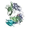
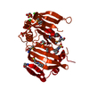
- PDB-4mjf: Crystal structure of a DUF4348 family protein (BVU_2238) from Bac... -
+
Open data
ID or keywords:
Loading...
-
Basic information
Entry
Database: PDB / ID: 4mjf
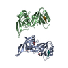
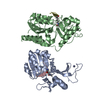
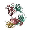
Title
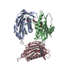
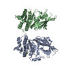
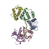
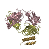
Crystal structure of a DUF4348 family protein (BVU_2238) from Bacteroides vulgatus ATCC 8482 at 1.99 A resolution
Components
hypothetical protein
Keywords
STRUCTURAL GENOMICS / UNKNOWN FUNCTION / PF14254 family / DUF4348 / Joint Center for Structural Genomics / JCSG / Protein Structure Initiative / PSI-BIOLOGY
Function / homology
Protein of unknown function DUF4348 / Domain of unknown function (DUF4348) / Prokaryotic membrane lipoprotein lipid attachment site profile. / DUF4348 domain-containing protein
Function and homology information
Biological species
Bacteroides vulgatus (bacteria)
Method
X-RAY DIFFRACTION / SYNCHROTRON / SAD / Resolution: 1.99 Å
Mass: 18.015 Da / Num. of mol.: 123 / Source method: isolated from a natural source / Formula: H2O
-
Details
Has protein modification
Y
Sequence details
THIS CONSTRUCT (25-286) WAS EXPRESSED WITH A PURIFICATION TAG MGSDKIHHHHHHENLYFQG. THE TAG WAS ...THIS CONSTRUCT (25-286) WAS EXPRESSED WITH A PURIFICATION TAG MGSDKIHHHHHHENLYFQG. THE TAG WAS REMOVED WITH TEV PROTEASE LEAVING ONLY A GLYCINE (0) FOLLOWED BY THE TARGET SEQUENCE.
-
Experimental details
-
Experiment
Experiment
Method: X-RAY DIFFRACTION / Number of used crystals: 1
-
Sample preparation
Crystal
Density Matthews: 2.43 Å3/Da / Density % sol: 49.3 %
Monochromator: single crystal Si(111) bent / Protocol: SAD / Monochromatic (M) / Laue (L): M / Scattering type: x-ray
Radiation wavelength
Wavelength: 0.97879 Å / Relative weight: 1
Reflection
Resolution: 1.99→48.085 Å / Num. obs: 20233 / % possible obs: 97.6 % / Observed criterion σ(I): -3 / Biso Wilson estimate: 44.225 Å2 / Rmerge F obs: 0.999 / Rmerge(I) obs: 0.037 / Rrim(I) all: 0.044 / Net I/σ(I): 15.21 / Num. measured all: 67280
Reflection shell
Diffraction-ID: 1
Resolution (Å)
Highest resolution (Å)
Rmerge F obs
Rmerge(I) obs
Mean I/σ(I) obs
Num. measured obs
Num. possible
Num. unique obs
Rrim(I) all
% possible all
1.99-2.06
0.728
0.562
2.1
6720
2020
1999
0.669
99
2.06-2.14
0.851
0.408
3
6789
1998
1976
0.484
98.9
2.14-2.24
0.924
0.273
4.3
6989
2117
2082
0.326
98.3
2.24-2.36
0.961
0.18
6.3
6599
2112
2054
0.217
97.3
2.36-2.51
0.986
0.113
9.8
7183
2097
2071
0.134
98.8
2.51-2.7
0.993
0.076
13.5
6617
2009
1958
0.09
97.5
2.7-2.97
0.997
0.049
19.1
6486
2052
1977
0.059
96.3
2.97-3.4
0.999
0.033
26.5
6853
2072
2025
0.039
97.7
3.4-4.27
0.999
0.025
33
6529
2064
1992
0.029
96.5
4.27
0.998
0.03
34.7
6515
2146
2059
0.036
95.9
-
Phasing
Phasing
Method: SAD
-
Processing
Software
Name
Version
Classification
NB
MolProbity
3beta29
modelbuilding
PDB_EXTRACT
3.1
dataextraction
SHELX
phasing
SHARP
phasing
XSCALE
datascaling
BUSTER-TNT
2.10.0
refinement
XDS
datareduction
SHELXD
phasing
BUSTER
2.10.0
refinement
Refinement
Method to determine structure: SAD / Resolution: 1.99→48.085 Å / Cor.coef. Fo:Fc: 0.9586 / Cor.coef. Fo:Fc free: 0.9526 / Occupancy max: 1 / Occupancy min: 0.3 / Cross valid method: THROUGHOUT / σ(F): 0 Details: 1.A MET-INHIBITION PROTOCOL WAS USED FOR SELENOMETHIONINE INCORPORATION DURING PROTEIN EXPRESSION. THE OCCUPANCY OF THE SE ATOMS IN THE MSE RESIDUES WAS REDUCED TO 0.75 FOR THE REDUCED ...Details: 1.A MET-INHIBITION PROTOCOL WAS USED FOR SELENOMETHIONINE INCORPORATION DURING PROTEIN EXPRESSION. THE OCCUPANCY OF THE SE ATOMS IN THE MSE RESIDUES WAS REDUCED TO 0.75 FOR THE REDUCED SCATTERING POWER DUE TO PARTIAL S-MET INCORPORATION. 2.ATOM RECORD CONTAINS SUM OF TLS AND RESIDUAL B FACTORS. 3.ANISOU RECORD CONTAINS SUM OF TLS AND RESIDUAL U FACTORS. 4.SULFATE (SO4), CAPS (CXS), CHLORIDE (CL) AND 1,2-ETHANEDIOL (EDO) FROM THE CRYSTALLIZATION AND CRYOPROTECTANT SOLUTION HAVE BEEN MODELED. 5.RESIDUES 173-174 ARE IN A REGION OF ELECTRON DENSITY THAT IS NOT WELL-DEFINED.
In the structure databanks used in Yorodumi, some data are registered as the other names, "COVID-19 virus" and "2019-nCoV". Here are the details of the virus and the list of structure data.
Jan 31, 2019. EMDB accession codes are about to change! (news from PDBe EMDB page)
EMDB accession codes are about to change! (news from PDBe EMDB page)
The allocation of 4 digits for EMDB accession codes will soon come to an end. Whilst these codes will remain in use, new EMDB accession codes will include an additional digit and will expand incrementally as the available range of codes is exhausted. The current 4-digit format prefixed with “EMD-” (i.e. EMD-XXXX) will advance to a 5-digit format (i.e. EMD-XXXXX), and so on. It is currently estimated that the 4-digit codes will be depleted around Spring 2019, at which point the 5-digit format will come into force.
The EM Navigator/Yorodumi systems omit the EMD- prefix.
Related info.:Q: What is EMD? / ID/Accession-code notation in Yorodumi/EM Navigator
Yorodumi is a browser for structure data from EMDB, PDB, SASBDB, etc.
This page is also the successor to EM Navigator detail page, and also detail information page/front-end page for Omokage search.
The word "yorodu" (or yorozu) is an old Japanese word meaning "ten thousand". "mi" (miru) is to see.
Related info.:EMDB / PDB / SASBDB / Comparison of 3 databanks / Yorodumi Search / Aug 31, 2016. New EM Navigator & Yorodumi / Yorodumi Papers / Jmol/JSmol / Function and homology information / Changes in new EM Navigator and Yorodumi
 Movie
Movie Controller

 Yorodumi
 Open data
 Basic information
 Components
 Keywords
 Function and homology information
 Bacteroides vulgatus (bacteria)
 X-RAY DIFFRACTION /
  Authors
 Citation
 Structure visualization
 Downloads & links
 Other downloads
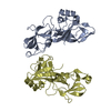

 PDBj
PDBj


 Assembly
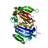


 Mass: 221.317 Da / Num. of mol.: 2 / Source method: obtained synthetically / Formula: C9H19NO3S / Comment: pH buffer*YM
 Mass: 96.063 Da / Num. of mol.: 4 / Source method: obtained synthetically / Formula: SO4
 Mass: 62.068 Da / Num. of mol.: 5 / Source method: obtained synthetically / Formula: C2H6O2
 Mass: 35.453 Da / Num. of mol.: 4 / Source method: obtained synthetically / Formula: Cl
 Sample preparation
 / Beamline: BL11-1 / Wavelength: 0.97879
  Processing