- PDB-2ps3: Structure and metal binding properties of ZnuA, a periplasmic zin... -
+
Open data
ID or keywords:
Loading...
-
Basic information
Entry
Database: PDB / ID: 2ps3
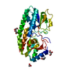
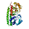
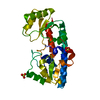
Title
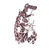
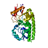
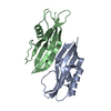
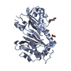
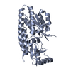
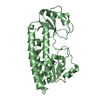
Structure and metal binding properties of ZnuA, a periplasmic zinc transporter from Escherichia coli
Components
High-affinity zinc uptake system protein znuA
Keywords
METAL TRANSPORT / The protein consists of two domains with a typical (beta/alfa)4 fold
Function / homology
Function and homology information
zinc ion import across plasma membrane / Metal ion assimilation from the host / zinc ion transport / ATP-binding cassette (ABC) transporter complex, substrate-binding subunit-containing / periplasmic space / cell adhesion / zinc ion binding / membrane Similarity search - Function
High-affinity zinc uptake system protein ZnuA / Adhesion lipoprotein / : / Periplasmic solute binding protein, ZnuA-like / Zinc-uptake complex component A periplasmic / Nitrogenase molybdenum iron protein domain / Rossmann fold / 3-Layer(aba) Sandwich / Alpha Beta Similarity search - Domain/homology
Mass: 31177.219 Da / Num. of mol.: 2 Source method: isolated from a genetically manipulated source Source: (gene. exp.) Escherichia coli (E. coli) / Production host: Escherichia coli (E. coli) / References: UniProt: P39172
Mass: 18.015 Da / Num. of mol.: 49 / Source method: isolated from a natural source / Formula: H2O
Has protein modification
Y
-
Experimental details
-
Experiment
Experiment
Method: X-RAY DIFFRACTION / Number of used crystals: 1
-
Sample preparation
Crystal
Density Matthews: 2.27 Å3/Da / Density % sol: 45.82 %
Crystal grow
Temperature: 293 K / Method: vapor diffusion, hanging drop / pH: 4.6 Details: 5 mg/mL protein in 20 mM MOPS, pH 7.0, 5 mM EDTA, 20 mM NaCl, 5 % glycerol was mixed 1:1 with a cryoprotectant solution containing 35.5 % (w/v) PEG 4000, 0.2 M ammonium acetate, 9 % ...Details: 5 mg/mL protein in 20 mM MOPS, pH 7.0, 5 mM EDTA, 20 mM NaCl, 5 % glycerol was mixed 1:1 with a cryoprotectant solution containing 35.5 % (w/v) PEG 4000, 0.2 M ammonium acetate, 9 % glycerol, 0.1 M sodium acetate, pH 4.6, VAPOR DIFFUSION, HANGING DROP, temperature 293K
-
Data collection
Diffraction
ID
Mean temperature (K)
Crystal-ID
1
100
1
2
1
3
1
Diffraction source
Source
Site
Beamline
ID
Wavelength
SYNCHROTRON
APS
5ID-B
1
0.97934
SYNCHROTRON
APS
19-ID
2
0.97934
SYNCHROTRON
APS
23-ID-D
3
0.97934
Detector
Type: MAR scanner 300 mm plate / Detector: IMAGE PLATE / Date: Jul 6, 2006 / Details: mirrors
Radiation
Monochromator: Si 111 CHANNEL / Protocol: SINGLE WAVELENGTH / Monochromatic (M) / Laue (L): M / Scattering type: x-ray
Radiation wavelength
Wavelength: 0.97934 Å / Relative weight: 1
Reflection
Resolution: 2.45→35.1 Å / Num. obs: 20560 / % possible obs: 98.7 % / Redundancy: 6.6 % / Biso Wilson estimate: 58.3 Å2 / Rsym value: 0.084
In the structure databanks used in Yorodumi, some data are registered as the other names, "COVID-19 virus" and "2019-nCoV". Here are the details of the virus and the list of structure data.
Jan 31, 2019. EMDB accession codes are about to change! (news from PDBe EMDB page)
EMDB accession codes are about to change! (news from PDBe EMDB page)
The allocation of 4 digits for EMDB accession codes will soon come to an end. Whilst these codes will remain in use, new EMDB accession codes will include an additional digit and will expand incrementally as the available range of codes is exhausted. The current 4-digit format prefixed with “EMD-” (i.e. EMD-XXXX) will advance to a 5-digit format (i.e. EMD-XXXXX), and so on. It is currently estimated that the 4-digit codes will be depleted around Spring 2019, at which point the 5-digit format will come into force.
The EM Navigator/Yorodumi systems omit the EMD- prefix.
Related info.:Q: What is EMD? / ID/Accession-code notation in Yorodumi/EM Navigator
Yorodumi is a browser for structure data from EMDB, PDB, SASBDB, etc.
This page is also the successor to EM Navigator detail page, and also detail information page/front-end page for Omokage search.
The word "yorodu" (or yorozu) is an old Japanese word meaning "ten thousand". "mi" (miru) is to see.
Related info.:EMDB / PDB / SASBDB / Comparison of 3 databanks / Yorodumi Search / Aug 31, 2016. New EM Navigator & Yorodumi / Yorodumi Papers / Jmol/JSmol / Function and homology information / Changes in new EM Navigator and Yorodumi
 Movie
Movie Controller

 Yorodumi
 Open data
 Basic information
 Components
 Keywords
 Function and homology information
 X-RAY DIFFRACTION /
  Authors
 Citation
 Structure visualization
 Downloads & links
 Other downloads


 PDBj
PDBj Assembly
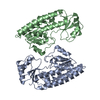
 Mass: 18.015 Da / Num. of mol.: 49 / Source method: isolated from a natural source / Formula: H2O
 Sample preparation
 Processing