- PDB-2osv: Crystal Structure of ZnuA from E. coli -
+
Open data
ID or keywords:
Loading...
-
Basic information
Entry
Database: PDB / ID: 2osv
Title
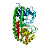
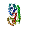
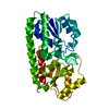
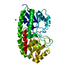
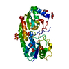
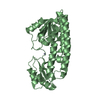
Crystal Structure of ZnuA from E. coli
Components
High-affinity zinc uptake system protein znuA
Keywords
METAL TRANSPORT / protein-zinc complex
Function / homology
Function and homology information
zinc ion import across plasma membrane / Metal ion assimilation from the host / zinc ion transport / ATP-binding cassette (ABC) transporter complex, substrate-binding subunit-containing / periplasmic space / cell adhesion / zinc ion binding / membrane Similarity search - Function
High-affinity zinc uptake system protein ZnuA / Adhesion lipoprotein / : / Periplasmic solute binding protein, ZnuA-like / Zinc-uptake complex component A periplasmic / Nitrogenase molybdenum iron protein domain / Rossmann fold / 3-Layer(aba) Sandwich / Alpha Beta Similarity search - Domain/homology
Journal: J.Mol.Biol. / Year: 2007 Title: Crystal Structure of the Zinc-binding Transport Protein ZnuA from Escherichia coli Reveals an Unexpected Variation in Metal Coordination. Authors: Li, H. / Jogl, G.
Mass: 31177.219 Da / Num. of mol.: 2 / Fragment: active form (N-terminal signal peptide cleaved) / Source method: isolated from a natural source / Source: (natural) Escherichia coli (E. coli) / Strain: BL21 Star / References: UniProt: P39172
In the structure databanks used in Yorodumi, some data are registered as the other names, "COVID-19 virus" and "2019-nCoV". Here are the details of the virus and the list of structure data.
Jan 31, 2019. EMDB accession codes are about to change! (news from PDBe EMDB page)
EMDB accession codes are about to change! (news from PDBe EMDB page)
The allocation of 4 digits for EMDB accession codes will soon come to an end. Whilst these codes will remain in use, new EMDB accession codes will include an additional digit and will expand incrementally as the available range of codes is exhausted. The current 4-digit format prefixed with “EMD-” (i.e. EMD-XXXX) will advance to a 5-digit format (i.e. EMD-XXXXX), and so on. It is currently estimated that the 4-digit codes will be depleted around Spring 2019, at which point the 5-digit format will come into force.
The EM Navigator/Yorodumi systems omit the EMD- prefix.
Related info.:Q: What is EMD? / ID/Accession-code notation in Yorodumi/EM Navigator
Yorodumi is a browser for structure data from EMDB, PDB, SASBDB, etc.
This page is also the successor to EM Navigator detail page, and also detail information page/front-end page for Omokage search.
The word "yorodu" (or yorozu) is an old Japanese word meaning "ten thousand". "mi" (miru) is to see.
Related info.:EMDB / PDB / SASBDB / Comparison of 3 databanks / Yorodumi Search / Aug 31, 2016. New EM Navigator & Yorodumi / Yorodumi Papers / Jmol/JSmol / Function and homology information / Changes in new EM Navigator and Yorodumi
 Movie
Movie Controller


 Open data
 Basic information
 Components
 Keywords
 Function and homology information
 X-RAY DIFFRACTION /
  Authors
 Citation
 Structure visualization
 Downloads & links
 Other downloads

 PDBj
PDBj Assembly
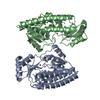


 Mass: 65.409 Da / Num. of mol.: 2 / Source method: obtained synthetically / Formula: Zn
 Mass: 18.015 Da / Num. of mol.: 596 / Source method: isolated from a natural source / Formula: H2O
 Sample preparation
 / Beamline: X4A / Wavelength: 0.979
  Processing