 Movie
Movie Controller


+ Open data

- Basic information
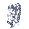
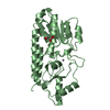
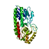
| Entry | Database: PDB / ID: 1k0f | ||||||
|---|---|---|---|---|---|---|---|
| Title | Crystal structure of Zn(II)-free T. pallidum TroA | ||||||
 Components Components | Periplasmic zinc-binding protein troA | ||||||
 Keywords Keywords | TRANSPORT PROTEIN / apo protein / helix backbone / closed conformation | ||||||
| Function / homology |  Function and homology information Function and homology informationzinc ion transport / periplasmic space / cell adhesion / metal ion binding Similarity search - Function | ||||||
| Biological species |  Treponema pallidum (bacteria) Treponema pallidum (bacteria) | ||||||
| Method |  X-RAY DIFFRACTION / MOLECULAR REPLACEMENT / Resolution: 2.5 Å X-RAY DIFFRACTION / MOLECULAR REPLACEMENT / Resolution: 2.5 Å | ||||||
 Authors Authors | Lee, Y.H. / Dorwart, M.R. / Hazlett, K.R. / Deka, R.K. / Norgard, M.V. / Radolf, J.D. / Hasemann, C.A. | ||||||
 Citation Citation | Journal: J.Bacteriol. / Year: 2002 Title: The crystal structure of Zn(II)-free Treponema pallidum TroA, a periplasmic metal-binding protein, reveals a closed conformation. Authors: Lee, Y.H. / Dorwart, M.R. / Hazlett, K.R. / Deka, R.K. / Norgard, M.V. / Radolf, J.D. / Hasemann, C.A. #1: Journal: Nat.Struct.Biol. / Year: 1999Title: Treponema pallidum TroA is a periplasmic zinc-binding protein with a helical backbone Authors: Lee, Y.H. / Deka, R.K. / Norgard, M.V. / Radolf, J.D. / Hasemann, C.A. | ||||||
| History |
|
- Structure visualization
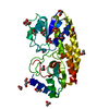
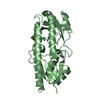
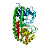
| Structure viewer | Molecule: MolmilJmol/JSmol |
|---|
- Downloads & links
-Download
| PDBx/mmCIF format | 1k0f.cif.gz | 65.3 KB | Display | PDBx/mmCIF format |
|---|---|---|---|---|
| PDB format | pdb1k0f.ent.gz | 48.2 KB | Display | PDB format |
| PDBx/mmJSON format | 1k0f.json.gz | Tree view | PDBx/mmJSON format | |
| Others |  Other downloads Other downloads |
-Validation report
| Arichive directory | https://data.pdbj.org/pub/pdb/validation_reports/k0/1k0fftp://data.pdbj.org/pub/pdb/validation_reports/k0/1k0f | HTTPS FTP |
|---|
-Related structure data
| Related structure data |  1toaS S: Starting model for refinement |
|---|---|
| Similar structure data |


-Links
 PDBj
PDBj- Assembly
| Deposited unit | 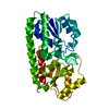
| ||||||||
|---|---|---|---|---|---|---|---|---|---|
| 1 |
| ||||||||
| Unit cell |
|
-Components
| #1: Protein | Mass: 30355.562 Da / Num. of mol.: 1 Source method: isolated from a genetically manipulated source Source: (gene. exp.) Treponema pallidum (bacteria) / Gene: troa / Plasmid: pProEx1 / Production host: |
|---|---|
| #2: Water | ChemComp-HOH /  Mass: 18.015 Da / Num. of mol.: 28 / Source method: isolated from a natural source / Formula: H2O Mass: 18.015 Da / Num. of mol.: 28 / Source method: isolated from a natural source / Formula: H2O |
-Experimental details
-Experiment
| Experiment | Method: X-RAY DIFFRACTION / Number of used crystals: 1 |
|---|
- Sample preparation
| Crystal | Density Matthews: 3.75 Å3/Da / Density % sol: 67.24 % | |||||||||||||||||||||||||||||||||||||||||||||||||||||||||||||||
|---|---|---|---|---|---|---|---|---|---|---|---|---|---|---|---|---|---|---|---|---|---|---|---|---|---|---|---|---|---|---|---|---|---|---|---|---|---|---|---|---|---|---|---|---|---|---|---|---|---|---|---|---|---|---|---|---|---|---|---|---|---|---|---|---|
| Crystal grow | Temperature: 298 K / Method: vapor diffusion, hanging drop / pH: 4.6 Details: PEG2000, pH 4.6, VAPOR DIFFUSION, HANGING DROP, temperature 298K | |||||||||||||||||||||||||||||||||||||||||||||||||||||||||||||||
| Crystal grow | *PLUS Temperature: 20 ℃ / pH: 7.4 | |||||||||||||||||||||||||||||||||||||||||||||||||||||||||||||||
| Components of the solutions | *PLUS
|
-Data collection
| Diffraction source | Source: ROTATING ANODE / Type: RIGAKU RU200 / Wavelength: 1.54 Å |
|---|---|
| Detector | Type: RIGAKU RAXIS IV / Detector: IMAGE PLATE / Date: Jan 10, 2000 / Details: Osmic confocal |
| Radiation | Monochromator: Graphite / Protocol: SINGLE WAVELENGTH / Monochromatic (M) / Laue (L): M / Scattering type: x-ray |
| Radiation wavelength | Wavelength: 1.54 Å / Relative weight: 1 |
| Reflection | Resolution: 2.5→30 Å / Num. all: 13646 / Num. obs: 13646 / % possible obs: 90.3 % / Observed criterion σ(F): 1 / Observed criterion σ(I): 1 / Redundancy: 3.4 % / Biso Wilson estimate: 56.4 Å2 / Rsym value: 0.078 / Net I/σ(I): 17.4 |
| Reflection shell | Highest resolution: 2.5 Å / Redundancy: 3.4 % / Rmerge(I) obs: 0.46 / Mean I/σ(I) obs: 2.5 / Num. unique all: 13646 / % possible all: 92.1 |
| Reflection | *PLUS Rmerge(I) obs: 0.078 |
| Reflection shell | *PLUS % possible obs: 92.1 % |
- Processing
| Software |
| ||||||||||||||||||||
|---|---|---|---|---|---|---|---|---|---|---|---|---|---|---|---|---|---|---|---|---|---|
| Refinement | Method to determine structure: MOLECULAR REPLACEMENT Starting model: pdb entry 1toa Resolution: 2.5→30 Å / Isotropic thermal model: Isotropic / Cross valid method: THROUGHOUT / σ(F): 0 / Stereochemistry target values: Engh & Huber Details: The structure factor file for this entry has been removed from the archive because it was found to be incomplete.
| ||||||||||||||||||||
| Displacement parameters | Biso mean: 36.7 Å2 | ||||||||||||||||||||
| Refinement step | Cycle: LAST / Resolution: 2.5→30 Å
| ||||||||||||||||||||
| Refine LS restraints |
| ||||||||||||||||||||
| Refinement | *PLUS Lowest resolution: 30 Å / σ(F): 0 / Rfactor obs: 0.208 | ||||||||||||||||||||
| Solvent computation | *PLUS | ||||||||||||||||||||
| Displacement parameters | *PLUS | ||||||||||||||||||||
| Refine LS restraints | *PLUS
|
