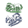
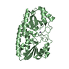
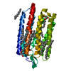
| Deposited unit | B: Periplasmic solute binding protein
A: Periplasmic solute binding protein
hetero molecules
| Theoretical mass | Number of molelcules |
|---|
| Total (without water) | 63,330 | 4 |
|---|
| Polymers | 63,199 | 2 |
|---|
| Non-polymers | 131 | 2 |
|---|
| Water | 1,027 | 57 |
|---|
|
|---|
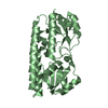
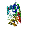
| 1 | B: Periplasmic solute binding protein
hetero molecules
| Theoretical mass | Number of molelcules |
|---|
| Total (without water) | 31,665 | 2 |
|---|
| Polymers | 31,600 | 1 |
|---|
| Non-polymers | 65 | 1 |
|---|
| Water | 18 | 1 |
|---|
| Type | Name | Symmetry operation | Number |
|---|
| identity operation | 1_555 | x,y,z | 1 |
|
|---|
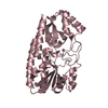
| 2 | A: Periplasmic solute binding protein
hetero molecules
| Theoretical mass | Number of molelcules |
|---|
| Total (without water) | 31,665 | 2 |
|---|
| Polymers | 31,600 | 1 |
|---|
| Non-polymers | 65 | 1 |
|---|
| Water | 18 | 1 |
|---|
| Type | Name | Symmetry operation | Number |
|---|
| identity operation | 1_555 | x,y,z | 1 |
|
|---|
| Unit cell | | Length a, b, c (Å) | 62.292, 105.042, 64.245 |
|---|
| Angle α, β, γ (deg.) | 90.000, 110.770, 90.000 |
|---|
| Int Tables number | 4 |
|---|
| Space group name H-M | P1211 |
|---|
|
|---|
| Noncrystallographic symmetry (NCS) | NCS domain: | ID | Ens-ID | Details (eV) |
|---|
| 1 | 1 | (chain A and (resid 26 through 35 or resid 37 through 80 or resid 82 through 309))| 2 | 1 | (chain B and (resid 26 through 35 or resid 37 through 80 or resid 82 through 309)) | |
NCS domain segments: Ens-ID: 1 | Dom-ID | Component-ID | Beg auth comp-ID | Beg label comp-ID | End auth comp-ID | End label comp-ID | Selection details | Auth asym-ID | Label asym-ID | Auth seq-ID | Label seq-ID |
|---|
| 1 | 1 | PROPROILEILE(chain A and (resid 26 through 35 or resid 37 through 80 or resid 82 through 309))AB| 26 - 35 | 26 - 35 | | 1 | 2 | GLYGLYLEULEU(chain A and (resid 26 through 35 or resid 37 through 80 or resid 82 through 309))AB| 37 - 80 | 37 - 80 | | 1 | 3 | ASNASNARGARG(chain A and (resid 26 through 35 or resid 37 through 80 or resid 82 through 309))AB| 82 - 309 | 82 - 301 | | 2 | 1 | PROPROILEILE(chain B and (resid 26 through 35 or resid 37 through 80 or resid 82 through 309))BA| 26 - 35 | 26 - 35 | | 2 | 2 | GLYGLYLEULEU| (chain B and (resid 26 through 35 or resid | | | | | | | | | | | | | | | | | | | | | | | | | | | | | | | | |
|
|---|
 Movie
Movie Controller


 Open data
 Basic information
 Components
 Keywords
 Function and homology information
 Paracoccus denitrificans (bacteria)
 X-RAY DIFFRACTION /
  Authors
 Citation
 Structure visualization
 Downloads & links
 Other downloads


 PDBj
PDBj Assembly