 Movie
Movie Controller

[English] 日本語
 Yorodumi
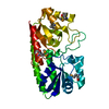
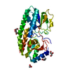
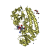
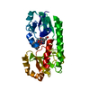
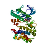
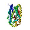
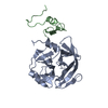
- PDB-6nsi: Crystal structure of Fe(III)-bound YtgA from Chlamydia trachomatis -
+ Open data

- Basic information
| Entry | Database: PDB / ID: 6nsi | ||||||||||||
|---|---|---|---|---|---|---|---|---|---|---|---|---|---|
| Title | Crystal structure of Fe(III)-bound YtgA from Chlamydia trachomatis | ||||||||||||
 Components Components | Manganese-binding protein | ||||||||||||
 Keywords Keywords | METAL BINDING PROTEIN / YtgA / solute-binding protein / Chlamydia trachomatis / iron acquisition / ABC transporter | ||||||||||||
| Function / homology |  Function and homology information Function and homology informationmetal ion transport / periplasmic space / cell adhesion / metal ion binding Similarity search - Function | ||||||||||||
| Biological species |   Chlamydia trachomatis (bacteria) Chlamydia trachomatis (bacteria) | ||||||||||||
| Method |  X-RAY DIFFRACTION / SYNCHROTRON / MOLECULAR REPLACEMENT / Resolution: 2.00006345565 Å X-RAY DIFFRACTION / SYNCHROTRON / MOLECULAR REPLACEMENT / Resolution: 2.00006345565 Å | ||||||||||||
 Authors Authors | Luo, Z. / Campbell, R. / Begg, S.L. / Kobe, B. / McDevitt, C.A. | ||||||||||||
| Funding support |  Australia, 3items Australia, 3items
| ||||||||||||
 Citation Citation | Journal: J.Bacteriol. / Year: 2019 Title: Structure and Metal Binding Properties of Chlamydia trachomatis YtgA. Authors: Luo, Z. / Neville, S.L. / Campbell, R. / Morey, J.R. / Menon, S. / Thomas, M. / Eijkelkamp, B.A. / Ween, M.P. / Huston, W.M. / Kobe, B. / McDevitt, C.A. | ||||||||||||
| History |
|
- Structure visualization
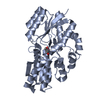
| Structure viewer | Molecule: MolmilJmol/JSmol |
|---|
- Downloads & links
-Download
| PDBx/mmCIF format | 6nsi.cif.gz | 138.7 KB | Display | PDBx/mmCIF format |
|---|---|---|---|---|
| PDB format | pdb6nsi.ent.gz | 101.6 KB | Display | PDB format |
| PDBx/mmJSON format | 6nsi.json.gz | Tree view | PDBx/mmJSON format | |
| Others |  Other downloads Other downloads |
-Validation report
| Arichive directory | https://data.pdbj.org/pub/pdb/validation_reports/ns/6nsiftp://data.pdbj.org/pub/pdb/validation_reports/ns/6nsi | HTTPS FTP |
|---|
-Related structure data
| Related structure data |  1pszS S: Starting model for refinement |
|---|---|
| Similar structure data |


-Links
 PDBj
PDBj- Assembly
| Deposited unit | 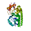
| ||||||||||||
|---|---|---|---|---|---|---|---|---|---|---|---|---|---|
| 1 |
| ||||||||||||
| Unit cell |
| ||||||||||||
| Components on special symmetry positions |
|
-Components
| #1: Protein | Mass: 32694.984 Da / Num. of mol.: 1 Source method: isolated from a genetically manipulated source Source: (gene. exp.) Chlamydia trachomatis (bacteria)Gene: troA, troA_1, ERS015772_00125, ERS082928_00414, ERS177788_00539 Production host: | ||||
|---|---|---|---|---|---|
| #2: Chemical | ChemComp-FE /   Mass: 55.845 Da / Num. of mol.: 1 / Source method: obtained synthetically / Formula: Fe Mass: 55.845 Da / Num. of mol.: 1 / Source method: obtained synthetically / Formula: Fe | ||||
| #3: Chemical |   Mass: 22.990 Da / Num. of mol.: 2 / Source method: obtained synthetically / Formula: Na Mass: 22.990 Da / Num. of mol.: 2 / Source method: obtained synthetically / Formula: Na#4: Chemical | ChemComp-CA / |   Mass: 40.078 Da / Num. of mol.: 1 / Source method: obtained synthetically / Formula: Ca Mass: 40.078 Da / Num. of mol.: 1 / Source method: obtained synthetically / Formula: Ca#5: Water | ChemComp-HOH / |  Mass: 18.015 Da / Num. of mol.: 208 / Source method: isolated from a natural source / Formula: H2O Mass: 18.015 Da / Num. of mol.: 208 / Source method: isolated from a natural source / Formula: H2O |
-Experimental details
-Experiment
| Experiment | Method: X-RAY DIFFRACTION / Number of used crystals: 1 |
|---|
- Sample preparation
| Crystal | Density Matthews: 2.35 Å3/Da / Density % sol: 47.69 % |
|---|---|
| Crystal grow | Temperature: 291 K / Method: vapor diffusion, hanging drop Details: 22 % (w/v) polyethylene glycol (PEG) 6000, 0.2 M CaCl2, 0.01 M FeCl3 and 0.1 M MES, pH 6.0 |
-Data collection
| Diffraction | Mean temperature: 100 K / Serial crystal experiment: N |
|---|---|
| Diffraction source | Source: SYNCHROTRON / Site: Australian Synchrotron / Beamline: MX1 / Wavelength: 0.954 Å |
| Detector | Type: ADSC QUANTUM 210r / Detector: CCD / Date: Jul 10, 2014 |
| Radiation | Protocol: SINGLE WAVELENGTH / Monochromatic (M) / Laue (L): M / Scattering type: x-ray |
| Radiation wavelength | Wavelength: 0.954 Å / Relative weight: 1 |
| Reflection | Resolution: 2→19.08 Å / Num. obs: 20476 / % possible obs: 100 % / Redundancy: 7.3 % / Biso Wilson estimate: 29.9512762463 Å2 / CC1/2: 0.99 / Rmerge(I) obs: 0.09 / Rpim(I) all: 0.04 / Net I/σ(I): 16.51 |
| Reflection shell | Resolution: 2→2.07 Å / Redundancy: 7.2 % / Rmerge(I) obs: 0.99 / Mean I/σ(I) obs: 2.03 / Num. unique obs: 2001 / CC1/2: 0.734 / Rpim(I) all: 0.39 / % possible all: 98.9 |
- Processing
| Software |
| ||||||||||||||||||||||||||||||||||||||||||||||||||||||||
|---|---|---|---|---|---|---|---|---|---|---|---|---|---|---|---|---|---|---|---|---|---|---|---|---|---|---|---|---|---|---|---|---|---|---|---|---|---|---|---|---|---|---|---|---|---|---|---|---|---|---|---|---|---|---|---|---|---|
| Refinement | Method to determine structure: MOLECULAR REPLACEMENT Starting model: 1PSZ Resolution: 2.00006345565→19.076725379 Å / SU ML: 0.269741598141 / Cross valid method: FREE R-VALUE / σ(F): 1.38024537978 / Phase error: 25.1403369776
| ||||||||||||||||||||||||||||||||||||||||||||||||||||||||
| Solvent computation | Shrinkage radii: 0.9 Å / VDW probe radii: 1.11 Å | ||||||||||||||||||||||||||||||||||||||||||||||||||||||||
| Displacement parameters | Biso mean: 39.1926758415 Å2 | ||||||||||||||||||||||||||||||||||||||||||||||||||||||||
| Refinement step | Cycle: LAST / Resolution: 2.00006345565→19.076725379 Å
| ||||||||||||||||||||||||||||||||||||||||||||||||||||||||
| Refine LS restraints |
| ||||||||||||||||||||||||||||||||||||||||||||||||||||||||
| LS refinement shell |
|
