 Movie
Movie Controller


+ Open data

- Basic information
| Entry | Database: PDB / ID: 1psz | ||||||
|---|---|---|---|---|---|---|---|
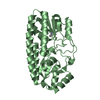
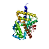
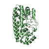
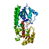
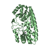
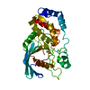
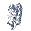
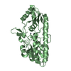
| Title | PNEUMOCOCCAL SURFACE ANTIGEN PSAA | ||||||
 Components Components | PROTEIN (SURFACE ANTIGEN PSAA) | ||||||
 Keywords Keywords | IMMUNE SYSTEM / PSAA / ABC-TYPE BINDING PROTEIN / METAL-BINDING PROTEIN / PNEUMOCOCCAL SURFACE ANTIGEN | ||||||
| Function / homology |  Function and homology information Function and homology informationmetal ion transport / cell adhesion / metal ion binding / plasma membrane Similarity search - Function | ||||||
| Biological species |   Streptococcus pneumoniae (bacteria) Streptococcus pneumoniae (bacteria) | ||||||
| Method |  X-RAY DIFFRACTION / MIR / Resolution: 2 Å X-RAY DIFFRACTION / MIR / Resolution: 2 Å | ||||||
 Authors Authors | Lawrence, M.C. / Pilling, P.A. / Epa, V.C. / Berry, A.M. / Ogunniyi, A.D. / Paton, J.C. | ||||||
 Citation Citation | Journal: Structure / Year: 1998 Title: The crystal structure of pneumococcal surface antigen PsaA reveals a metal-binding site and a novel structure for a putative ABC-type binding protein. Authors: Lawrence, M.C. / Pilling, P.A. / Epa, V.C. / Berry, A.M. / Ogunniyi, A.D. / Paton, J.C. #1: Journal: Acta Crystallogr.,Sect.D / Year: 1998Title: Expression, Purification and Preliminary X-Ray Crystallographic Analysis of PsaA, a Putative Metal-Transporter Protein of Streptococcus Pnuemoniae Authors: Pilling, P.A. / Lawrence, M.C. / Berry, A.M. / Ogunniyi, A.D. / Lock, R.A. / Paton, J.C. | ||||||
| History |
|
- Structure visualization
| Structure viewer | Molecule: MolmilJmol/JSmol |
|---|
- Downloads & links
-Download
| PDBx/mmCIF format | 1psz.cif.gz | 74.9 KB | Display | PDBx/mmCIF format |
|---|---|---|---|---|
| PDB format | pdb1psz.ent.gz | 55 KB | Display | PDB format |
| PDBx/mmJSON format | 1psz.json.gz | Tree view | PDBx/mmJSON format | |
| Others |  Other downloads Other downloads |
-Validation report
| Arichive directory | https://data.pdbj.org/pub/pdb/validation_reports/ps/1pszftp://data.pdbj.org/pub/pdb/validation_reports/ps/1psz | HTTPS FTP |
|---|
-Related structure data
| Similar structure data |
|---|


-Links
 PDBj
PDBj


- Assembly
| Deposited unit | 
| ||||||||
|---|---|---|---|---|---|---|---|---|---|
| 1 |
| ||||||||
| Unit cell |
|
-Components
| #1: Protein | Mass: 34143.254 Da / Num. of mol.: 1 Mutation: INCLUDES N-TERMINAL PURIFICATION TAG WRGSHHHHHHGSA Source method: isolated from a genetically manipulated source Details: THE EXPRESSED CONSTRUCT LACKS THE PRO-LIPOPROTEIN RECOGNITION SEQUENCE LXXC Source: (gene. exp.) Streptococcus pneumoniae (bacteria) / Strain: D39 (SEROTYPE 2) / Cellular location: EXTRACELLULAR / Cellular location (production host): CYTOPLASM / Production host: |
|---|---|
| #2: Chemical | ChemComp-ZN /   Mass: 65.409 Da / Num. of mol.: 1 / Source method: obtained synthetically / Formula: Zn Mass: 65.409 Da / Num. of mol.: 1 / Source method: obtained synthetically / Formula: Zn |
| #3: Water | ChemComp-HOH /  Mass: 18.015 Da / Num. of mol.: 225 / Source method: isolated from a natural source / Formula: H2O Mass: 18.015 Da / Num. of mol.: 225 / Source method: isolated from a natural source / Formula: H2O |
| Sequence details | THE DATABASE ENTRY HAS RESIDUES 1 TO 309 THE CRYSTALLIZED PROTEIN HAS RESIDUES 20 TO 309 PRECEDED ...THE DATABASE ENTRY HAS RESIDUES 1 TO 309 THE CRYSTALLIZ |
-Experimental details
-Experiment
| Experiment | Method: X-RAY DIFFRACTION / Number of used crystals: 1 |
|---|
- Sample preparation
| Crystal | Density Matthews: 2.05 Å3/Da / Density % sol: 40 % | |||||||||||||||||||||||||||||||||||||||||||||||||
|---|---|---|---|---|---|---|---|---|---|---|---|---|---|---|---|---|---|---|---|---|---|---|---|---|---|---|---|---|---|---|---|---|---|---|---|---|---|---|---|---|---|---|---|---|---|---|---|---|---|---|
| Crystal grow | pH: 7.5 Details: CRYSTALS WERE GROWN BY MICRO OR MACROSEEDING IN HANGING DROPS. PROTEIN CONCENTRATION 18 MG/ML. PRECIPITANT 3.0 M K2HPO4/NAH2PO4 (PH 7.5) 0.1 M MES ( PH 6.5) 0.1 M GUHCL AT 18 DEGREES. | |||||||||||||||||||||||||||||||||||||||||||||||||
| Crystal grow | *PLUS Temperature: 291 K / Method: vapor diffusion, hanging dropDetails: Pilling, P.A., (1998) Acta Crystallogr., Sect.D, 54, 1464. | |||||||||||||||||||||||||||||||||||||||||||||||||
| Components of the solutions | *PLUS
|
-Data collection
| Diffraction | Mean temperature: 108 K |
|---|---|
| Diffraction source | Source: ROTATING ANODE / Type: MACSCIENCE M18X / Wavelength: 1.5418 |
| Detector | Type: RIGAKU RAXIS IV / Detector: IMAGE PLATE / Date: Jun 15, 1997 / Details: MIRRORS |
| Radiation | Protocol: SINGLE WAVELENGTH / Monochromatic (M) / Laue (L): M / Scattering type: x-ray |
| Radiation wavelength | Wavelength: 1.5418 Å / Relative weight: 1 |
| Reflection | Resolution: 2→20 Å / Num. obs: 19354 / % possible obs: 99.5 % / Redundancy: 3.7 % / Rmerge(I) obs: 0.11 / Net I/σ(I): 12.4 |
| Reflection shell | Resolution: 2→2.05 Å / Redundancy: 3.5 % / Rmerge(I) obs: 0.39 / Mean I/σ(I) obs: 3.4 / % possible all: 99 |
| Reflection shell | *PLUS % possible obs: 99.6 % |
- Processing
| Software |
| ||||||||||||||||||||||||||||||||||||||||||||||||||||||||||||||||||||||||||||||||||||
|---|---|---|---|---|---|---|---|---|---|---|---|---|---|---|---|---|---|---|---|---|---|---|---|---|---|---|---|---|---|---|---|---|---|---|---|---|---|---|---|---|---|---|---|---|---|---|---|---|---|---|---|---|---|---|---|---|---|---|---|---|---|---|---|---|---|---|---|---|---|---|---|---|---|---|---|---|---|---|---|---|---|---|---|---|---|
| Refinement | Method to determine structure: MIR / Resolution: 2→8 Å / SU B: 3.9 / SU ML: 0.11 / Cross valid method: THROUGHOUT / σ(F): 0 / ESU R: 0.2 / ESU R Free: 0.18 Details: NO STEREOCHEMICAL CONSTRAINTS WERE APPLIED TO THE ZINC ION DURING REFINEMENT
| ||||||||||||||||||||||||||||||||||||||||||||||||||||||||||||||||||||||||||||||||||||
| Displacement parameters | Biso mean: 17.3 Å2 | ||||||||||||||||||||||||||||||||||||||||||||||||||||||||||||||||||||||||||||||||||||
| Refinement step | Cycle: LAST / Resolution: 2→8 Å
| ||||||||||||||||||||||||||||||||||||||||||||||||||||||||||||||||||||||||||||||||||||
| Refine LS restraints |
| ||||||||||||||||||||||||||||||||||||||||||||||||||||||||||||||||||||||||||||||||||||
| Software | *PLUS Name: REFMAC / Classification: refinement | ||||||||||||||||||||||||||||||||||||||||||||||||||||||||||||||||||||||||||||||||||||
| Refinement | *PLUS Highest resolution: 2 Å / σ(F): 0 / % reflection Rfree: 5 % / Rfactor obs: 0.178 | ||||||||||||||||||||||||||||||||||||||||||||||||||||||||||||||||||||||||||||||||||||
| Solvent computation | *PLUS | ||||||||||||||||||||||||||||||||||||||||||||||||||||||||||||||||||||||||||||||||||||
| Displacement parameters | *PLUS Biso mean: 17.3 Å2 | ||||||||||||||||||||||||||||||||||||||||||||||||||||||||||||||||||||||||||||||||||||
| Refine LS restraints | *PLUS Type: p_mcbond_it / Dev ideal target: 2 |
