 Movie
Movie Controller


+ Open data

- Basic information
| Entry | Database: PDB / ID: 4hr5 | ||||||
|---|---|---|---|---|---|---|---|
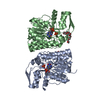
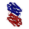
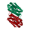
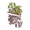
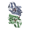
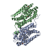
| Title | R2-like ligand-binding oxidase without metal cofactor | ||||||
 Components Components | Ribonuleotide reductase small subunit | ||||||
 Keywords Keywords | OXIDOREDUCTASE / R2-like ligand-binding oxidase / heterodinuclear Mn/Fe cofactor / ribonucleotide reductase R2 subunit fold / metalloprotein / manganese / iron / fatty acid/long-chain hydrocarbon ligand | ||||||
| Function / homology |  Function and homology information Function and homology informationdeoxyribonucleotide biosynthetic process / oxidoreductase activity / metal ion binding Similarity search - Function | ||||||
| Biological species |  Geobacillus kaustophilus (bacteria) Geobacillus kaustophilus (bacteria) | ||||||
| Method |  X-RAY DIFFRACTION / SYNCHROTRON / FOURIER SYNTHESIS / Resolution: 2.291 Å X-RAY DIFFRACTION / SYNCHROTRON / FOURIER SYNTHESIS / Resolution: 2.291 Å | ||||||
 Authors Authors | Griese, J.J. / Hogbom, M. | ||||||
 Citation Citation | Journal: Proc.Natl.Acad.Sci.USA / Year: 2013 Title: Direct observation of structurally encoded metal discrimination and ether bond formation in a heterodinuclear metalloprotein Authors: Griese, J.J. / Roos, K. / Cox, N. / Shafaat, H.S. / Branca, R.M.M. / Lehtio, J. / Graslund, A. / Lubitz, W. / Siegbahn, P.E.M. / Hogbom, M. | ||||||
| History |
|
- Structure visualization
| Structure viewer | Molecule: MolmilJmol/JSmol |
|---|
- Downloads & links
-Download
| PDBx/mmCIF format | 4hr5.cif.gz | 116.9 KB | Display | PDBx/mmCIF format |
|---|---|---|---|---|
| PDB format | pdb4hr5.ent.gz | 92.5 KB | Display | PDB format |
| PDBx/mmJSON format | 4hr5.json.gz | Tree view | PDBx/mmJSON format | |
| Others |  Other downloads Other downloads |
-Validation report
| Arichive directory | https://data.pdbj.org/pub/pdb/validation_reports/hr/4hr5ftp://data.pdbj.org/pub/pdb/validation_reports/hr/4hr5 | HTTPS FTP |
|---|
-Related structure data


-Links
 PDBj
PDBj


- Assembly
| Deposited unit | 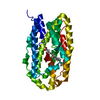
| |||||||||
|---|---|---|---|---|---|---|---|---|---|---|
| 1 | 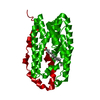
| |||||||||
| Unit cell |
| |||||||||
| Components on special symmetry positions |
|
-Components
| #1: Protein | Mass: 36962.809 Da / Num. of mol.: 1 Source method: isolated from a genetically manipulated source Source: (gene. exp.) Geobacillus kaustophilus (bacteria) / Strain: HTA246 / Gene: GK2771 / Plasmid: pET-46 Ek/LIC / Production host: References: UniProt: Q5KW80, ribonucleoside-diphosphate reductase |
|---|---|
| #2: Chemical | ChemComp-PLM /   Mass: 256.424 Da / Num. of mol.: 1 / Source method: obtained synthetically / Formula: C16H32O2 Mass: 256.424 Da / Num. of mol.: 1 / Source method: obtained synthetically / Formula: C16H32O2 |
| #3: Water | ChemComp-HOH /  Mass: 18.015 Da / Num. of mol.: 23 / Source method: isolated from a natural source / Formula: H2O Mass: 18.015 Da / Num. of mol.: 23 / Source method: isolated from a natural source / Formula: H2O |
-Experimental details
-Experiment
| Experiment | Method: X-RAY DIFFRACTION / Number of used crystals: 1 |
|---|
- Sample preparation
| Crystal | Density Matthews: 2.38 Å3/Da / Density % sol: 48.26 % |
|---|---|
| Crystal grow | Temperature: 295 K / Method: vapor diffusion, hanging drop / pH: 7 Details: 17.5%(w/v) PEG 1500, 0.1M HEPES-Na, pH 7.0, VAPOR DIFFUSION, HANGING DROP, temperature 295K |
-Data collection
| Diffraction | Mean temperature: 100 K |
|---|---|
| Diffraction source | Source: SYNCHROTRON / Site: BESSY  / Beamline: 14.1 / Wavelength: 0.96 Å / Beamline: 14.1 / Wavelength: 0.96 Å |
| Detector | Type: RAYONIX MX-225 / Detector: CCD / Date: Jan 25, 2012 |
| Radiation | Monochromator: KMC-1 / Protocol: SINGLE WAVELENGTH / Monochromatic (M) / Laue (L): M / Scattering type: x-ray |
| Radiation wavelength | Wavelength: 0.96 Å / Relative weight: 1 |
| Reflection | Resolution: 2.29→30 Å / Num. all: 16220 / Num. obs: 16220 / % possible obs: 99.8 % / Observed criterion σ(F): 0 / Observed criterion σ(I): -3 / Redundancy: 14.6 % / Biso Wilson estimate: 54.307 Å2 / Rmerge(I) obs: 0.071 / Net I/σ(I): 25.87 |
| Reflection shell | Resolution: 2.29→2.43 Å / Redundancy: 14.7 % / Rmerge(I) obs: 0.754 / Mean I/σ(I) obs: 4.13 / Num. unique all: 2540 / % possible all: 99.1 |
- Processing
| Software |
| |||||||||||||||||||||||||||||||||||||||||||||||||
|---|---|---|---|---|---|---|---|---|---|---|---|---|---|---|---|---|---|---|---|---|---|---|---|---|---|---|---|---|---|---|---|---|---|---|---|---|---|---|---|---|---|---|---|---|---|---|---|---|---|---|
| Refinement | Method to determine structure: FOURIER SYNTHESIS / Resolution: 2.291→28.378 Å / SU ML: 0.22 / Isotropic thermal model: isotropic / Cross valid method: THROUGHOUT / σ(F): 1.99 / Phase error: 26.12 / Stereochemistry target values: ML
| |||||||||||||||||||||||||||||||||||||||||||||||||
| Solvent computation | Shrinkage radii: 0.9 Å / VDW probe radii: 1.11 Å / Solvent model: FLAT BULK SOLVENT MODEL | |||||||||||||||||||||||||||||||||||||||||||||||||
| Displacement parameters | Biso mean: 71.4 Å2 | |||||||||||||||||||||||||||||||||||||||||||||||||
| Refinement step | Cycle: LAST / Resolution: 2.291→28.378 Å
| |||||||||||||||||||||||||||||||||||||||||||||||||
| Refine LS restraints |
| |||||||||||||||||||||||||||||||||||||||||||||||||
| LS refinement shell | Refine-ID: X-RAY DIFFRACTION / Total num. of bins used: 6
|
