 Movie
Movie Controller

[English] 日本語
 Yorodumi
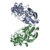
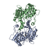
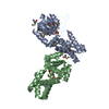
- PDB-6vpi: TPX2 residues 7-20 fused to Aurora A residues 116-389 C247V + D25... -
+ Open data

- Basic information
| Entry | Database: PDB / ID: 6vpi | |||||||||||||||
|---|---|---|---|---|---|---|---|---|---|---|---|---|---|---|---|---|
| Title | TPX2 residues 7-20 fused to Aurora A residues 116-389 C247V + D256N + C319V triple mutant disulfide homodimer in complex with AMP-PNP | |||||||||||||||
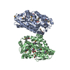
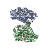
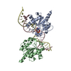
 Components Components | TPX2 fragment - Aurora A kinase domain fusion | |||||||||||||||
 Keywords Keywords | TRANSFERASE / Ser/Thr kinase | |||||||||||||||
| Function / homology |  Function and homology information Function and homology informationactivation of protein kinase activity / negative regulation of microtubule depolymerization / Interaction between PHLDA1 and AURKA / regulation of centrosome cycle / axon hillock / spindle assembly involved in female meiosis I / cilium disassembly / microtubule nucleation / spindle pole centrosome / importin-alpha family protein binding ...activation of protein kinase activity / negative regulation of microtubule depolymerization / Interaction between PHLDA1 and AURKA / regulation of centrosome cycle / axon hillock / spindle assembly involved in female meiosis I / cilium disassembly / microtubule nucleation / spindle pole centrosome / importin-alpha family protein binding / mitotic centrosome separation / histone H3S10 kinase activity / chromosome passenger complex / positive regulation of oocyte maturation / anterior/posterior axis specification / protein localization to centrosome / pronucleus / germinal vesicle / meiotic spindle / centrosome localization / neuron projection extension / spindle organization / positive regulation of mitochondrial fission / mitotic spindle pole / intercellular bridge / spindle midzone / SUMOylation of DNA replication proteins / mitotic spindle assembly / negative regulation of protein binding / regulation of G2/M transition of mitotic cell cycle / positive regulation of mitotic cell cycle / positive regulation of mitotic nuclear division / protein serine/threonine/tyrosine kinase activity / centriole / liver regeneration / TP53 Regulates Transcription of Genes Involved in G2 Cell Cycle Arrest / AURKA Activation by TPX2 / regulation of signal transduction by p53 class mediator / molecular function activator activity / mitotic spindle organization / protein serine/threonine kinase activator activity / regulation of cytokinesis / regulation of mitotic spindle organization / peptidyl-serine phosphorylation / regulation of protein stability / response to wounding / APC/C:Cdh1 mediated degradation of Cdc20 and other APC/C:Cdh1 targeted proteins in late mitosis/early G1 / FBXL7 down-regulates AURKA during mitotic entry and in early mitosis / G2/M transition of mitotic cell cycle / mitotic spindle / spindle / kinetochore / spindle pole / microtubule cytoskeleton / Regulation of PLK1 Activity at G2/M Transition / mitotic cell cycle / protein autophosphorylation / positive regulation of proteasomal ubiquitin-dependent protein catabolic process / midbody / Regulation of TP53 Activity through Phosphorylation / molecular adaptor activity / ciliary basal body / basolateral plasma membrane / microtubule / proteasome-mediated ubiquitin-dependent protein catabolic process / protein phosphorylation / protein kinase activity / non-specific serine/threonine protein kinase / postsynaptic density / protein heterodimerization activity / negative regulation of gene expression / protein serine kinase activity / cell division / protein serine/threonine kinase activity / centrosome / ubiquitin protein ligase binding / apoptotic process / negative regulation of apoptotic process / protein kinase binding / perinuclear region of cytoplasm / glutamatergic synapse / nucleoplasm / ATP binding / nucleus / cytosol Similarity search - Function | |||||||||||||||
| Biological species |  Homo sapiens (human) Homo sapiens (human) | |||||||||||||||
| Method |  X-RAY DIFFRACTION / MOLECULAR REPLACEMENT / molecular replacement / Resolution: 2 Å X-RAY DIFFRACTION / MOLECULAR REPLACEMENT / molecular replacement / Resolution: 2 Å | |||||||||||||||
 Authors Authors | Lim, D.C. / Yaffe, M.B. | |||||||||||||||
| Funding support |  United States, 4items United States, 4items
| |||||||||||||||
 Citation Citation | Journal: Sci.Signal. / Year: 2020 Title: Redox priming promotes Aurora A activation during mitosis. Authors: Lim, D.C. / Joukov, V. / Rettenmaier, T.J. / Kumagai, A. / Dunphy, W.G. / Wells, J.A. / Yaffe, M.B. | |||||||||||||||
| History |
|
- Structure visualization
| Structure viewer | Molecule: MolmilJmol/JSmol |
|---|
- Downloads & links
-Download
| PDBx/mmCIF format | 6vpi.cif.gz | 140 KB | Display | PDBx/mmCIF format |
|---|---|---|---|---|
| PDB format | pdb6vpi.ent.gz | 106.9 KB | Display | PDB format |
| PDBx/mmJSON format | 6vpi.json.gz | Tree view | PDBx/mmJSON format | |
| Others |  Other downloads Other downloads |
-Validation report
| Arichive directory | https://data.pdbj.org/pub/pdb/validation_reports/vp/6vpiftp://data.pdbj.org/pub/pdb/validation_reports/vp/6vpi | HTTPS FTP |
|---|
-Related structure data
| Related structure data | 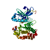 6vpgC 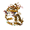 6vphC 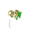 6vpjC  6vplC  6vpmC 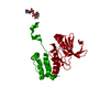 6xkaC 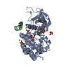 1ol5S S: Starting model for refinement C: citing same article ( |
|---|---|
| Similar structure data |


-Links
 PDBj
PDBj


- Assembly
| Deposited unit | 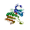
| ||||||||
|---|---|---|---|---|---|---|---|---|---|
| 1 | 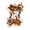
| ||||||||
| Unit cell |
| ||||||||
| Components on special symmetry positions |
|
-Components
| #1: Protein | Mass: 33897.742 Da / Num. of mol.: 1 / Fragment: kinase domain Source method: isolated from a genetically manipulated source Source: (gene. exp.) Homo sapiens (human)Gene: TPX2, C20orf1, C20orf2, DIL2, HCA519, AURKA, AIK, AIRK1, ARK1, AURA, AYK1, BTAK, IAK1, STK15, STK6 Plasmid: pET28a / Production host:  References: UniProt: Q9ULW0, UniProt: O14965, non-specific serine/threonine protein kinase | ||||||||
|---|---|---|---|---|---|---|---|---|---|
| #2: Chemical | ChemComp-ANP /   Mass: 506.196 Da / Num. of mol.: 1 / Source method: obtained synthetically / Formula: C10H17N6O12P3 / Feature type: SUBJECT OF INVESTIGATION / Comment: AMP-PNP, energy-carrying molecule analogue*YM Mass: 506.196 Da / Num. of mol.: 1 / Source method: obtained synthetically / Formula: C10H17N6O12P3 / Feature type: SUBJECT OF INVESTIGATION / Comment: AMP-PNP, energy-carrying molecule analogue*YM | ||||||||
| #3: Chemical |   Mass: 24.305 Da / Num. of mol.: 2 / Source method: obtained synthetically / Formula: Mg / Feature type: SUBJECT OF INVESTIGATION Mass: 24.305 Da / Num. of mol.: 2 / Source method: obtained synthetically / Formula: Mg / Feature type: SUBJECT OF INVESTIGATION#4: Chemical | ChemComp-MLA / |   Mass: 104.061 Da / Num. of mol.: 1 Mass: 104.061 Da / Num. of mol.: 1Source method: isolated from a genetically manipulated source Formula: C3H4O4 #5: Water | ChemComp-HOH / |  Mass: 18.015 Da / Num. of mol.: 233 / Source method: isolated from a natural source / Formula: H2O Mass: 18.015 Da / Num. of mol.: 233 / Source method: isolated from a natural source / Formula: H2OHas ligand of interest | Y | Has protein modification | Y | |
-Experimental details
-Experiment
| Experiment | Method: X-RAY DIFFRACTION / Number of used crystals: 1 |
|---|
- Sample preparation
| Crystal | Density Matthews: 3.44 Å3/Da / Density % sol: 64.25 % / Mosaicity: 0.359 ° |
|---|---|
| Crystal grow | Temperature: 298 K / Method: evaporation / pH: 7 Details: 0.336 M sodium malonate pH 7.5, 0.348 M sodium malonate pH 6.5 |
-Data collection
| Diffraction | Mean temperature: 100 K / Serial crystal experiment: N | |||||||||||||||||||||||||||||||||||||||||||||||||||||||||||||||||||||||||||||||||||||||||||||||||||||||||||||||||||||||||||||||||||||||||||||||||||||||||||||||||||||||||||||||||||||||||||||
|---|---|---|---|---|---|---|---|---|---|---|---|---|---|---|---|---|---|---|---|---|---|---|---|---|---|---|---|---|---|---|---|---|---|---|---|---|---|---|---|---|---|---|---|---|---|---|---|---|---|---|---|---|---|---|---|---|---|---|---|---|---|---|---|---|---|---|---|---|---|---|---|---|---|---|---|---|---|---|---|---|---|---|---|---|---|---|---|---|---|---|---|---|---|---|---|---|---|---|---|---|---|---|---|---|---|---|---|---|---|---|---|---|---|---|---|---|---|---|---|---|---|---|---|---|---|---|---|---|---|---|---|---|---|---|---|---|---|---|---|---|---|---|---|---|---|---|---|---|---|---|---|---|---|---|---|---|---|---|---|---|---|---|---|---|---|---|---|---|---|---|---|---|---|---|---|---|---|---|---|---|---|---|---|---|---|---|---|---|---|---|
| Diffraction source | Source: ROTATING ANODE / Type: RIGAKU / Wavelength: 1.5418 Å | |||||||||||||||||||||||||||||||||||||||||||||||||||||||||||||||||||||||||||||||||||||||||||||||||||||||||||||||||||||||||||||||||||||||||||||||||||||||||||||||||||||||||||||||||||||||||||||
| Detector | Type: RIGAKU SATURN 944 / Detector: CCD / Date: Apr 16, 2015 / Details: mirrors | |||||||||||||||||||||||||||||||||||||||||||||||||||||||||||||||||||||||||||||||||||||||||||||||||||||||||||||||||||||||||||||||||||||||||||||||||||||||||||||||||||||||||||||||||||||||||||||
| Radiation | Monochromator: mirrors / Protocol: SINGLE WAVELENGTH / Monochromatic (M) / Laue (L): M / Scattering type: x-ray | |||||||||||||||||||||||||||||||||||||||||||||||||||||||||||||||||||||||||||||||||||||||||||||||||||||||||||||||||||||||||||||||||||||||||||||||||||||||||||||||||||||||||||||||||||||||||||||
| Radiation wavelength | Wavelength: 1.5418 Å / Relative weight: 1 | |||||||||||||||||||||||||||||||||||||||||||||||||||||||||||||||||||||||||||||||||||||||||||||||||||||||||||||||||||||||||||||||||||||||||||||||||||||||||||||||||||||||||||||||||||||||||||||
| Reflection | Resolution: 2→23.33 Å / Num. obs: 32094 / % possible obs: 98.1 % / Redundancy: 3.9 % / Rmerge(I) obs: 0.047 / Rpim(I) all: 0.026 / Rrim(I) all: 0.054 / Χ2: 0.988 / Net I/σ(I): 13.9 / Num. measured all: 126757 | |||||||||||||||||||||||||||||||||||||||||||||||||||||||||||||||||||||||||||||||||||||||||||||||||||||||||||||||||||||||||||||||||||||||||||||||||||||||||||||||||||||||||||||||||||||||||||||
| Reflection shell | Diffraction-ID: 1
|
-Phasing
| Phasing | Method: molecular replacement |
|---|
- Processing
| Software |
| |||||||||||||||||||||||||||||||||||||||||||||||||||||||||||||||||||||||||||||||||||||||||||||||||||||||||||||||||||||||||||||
|---|---|---|---|---|---|---|---|---|---|---|---|---|---|---|---|---|---|---|---|---|---|---|---|---|---|---|---|---|---|---|---|---|---|---|---|---|---|---|---|---|---|---|---|---|---|---|---|---|---|---|---|---|---|---|---|---|---|---|---|---|---|---|---|---|---|---|---|---|---|---|---|---|---|---|---|---|---|---|---|---|---|---|---|---|---|---|---|---|---|---|---|---|---|---|---|---|---|---|---|---|---|---|---|---|---|---|---|---|---|---|---|---|---|---|---|---|---|---|---|---|---|---|---|---|---|---|
| Refinement | Method to determine structure: MOLECULAR REPLACEMENT Starting model: 1OL5 Resolution: 2→23.26 Å / SU ML: 0.19 / Cross valid method: THROUGHOUT / σ(F): 1.33 / Phase error: 22.25
| |||||||||||||||||||||||||||||||||||||||||||||||||||||||||||||||||||||||||||||||||||||||||||||||||||||||||||||||||||||||||||||
| Solvent computation | Shrinkage radii: 0.9 Å / VDW probe radii: 1.11 Å | |||||||||||||||||||||||||||||||||||||||||||||||||||||||||||||||||||||||||||||||||||||||||||||||||||||||||||||||||||||||||||||
| Displacement parameters | Biso max: 120.3 Å2 / Biso mean: 41.7298 Å2 / Biso min: 20.54 Å2 | |||||||||||||||||||||||||||||||||||||||||||||||||||||||||||||||||||||||||||||||||||||||||||||||||||||||||||||||||||||||||||||
| Refinement step | Cycle: final / Resolution: 2→23.26 Å
| |||||||||||||||||||||||||||||||||||||||||||||||||||||||||||||||||||||||||||||||||||||||||||||||||||||||||||||||||||||||||||||
| LS refinement shell | Refine-ID: X-RAY DIFFRACTION / Rfactor Rfree error: 0 / Total num. of bins used: 14
| |||||||||||||||||||||||||||||||||||||||||||||||||||||||||||||||||||||||||||||||||||||||||||||||||||||||||||||||||||||||||||||
| Refinement TLS params. | Method: refined / Refine-ID: X-RAY DIFFRACTION
| |||||||||||||||||||||||||||||||||||||||||||||||||||||||||||||||||||||||||||||||||||||||||||||||||||||||||||||||||||||||||||||
| Refinement TLS group |
|
