- PDB-4lqx: Crystal structure of a TENA/THI-4 domain-containing protein (SSO2... -
+
Open data
ID or keywords:
Loading...
-
Basic information
Entry
Database: PDB / ID: 4lqx
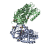
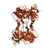
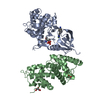
Title
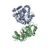
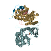
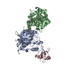
Crystal structure of a TENA/THI-4 domain-containing protein (SSO2700) from Sulfolobus solfataricus P2 at 2.34 A resolution
Components
TENA/THI-4 domain-containing protein
Keywords
OXIDOREDUCTASE / Two domains protein / N-terminal is prokaryotic zinc finger domain and C-terminal is TENA_THI-4 domain (PF03070) / Structural Genomics / Joint Center for Structural Genomics / JCSG / Protein Structure Initiative / PSI-BIOLOGY
Function / homology
Function and homology information
: / pyrimidine-containing compound metabolic process / sulfur compound metabolic process / small molecule metabolic process / oxidoreductase activity / metal ion binding Similarity search - Function
Uncharacterised conserved protein UCP032676, PqqC / UCP032676, C2H2 type zinc-finger domain / C2H2 type zinc-finger (1 copy) / Pyrroloquinoline-quinone synthase-like / Thiaminase-2/PQQC / TENA/THI-4/PQQC family / Heme oxygenase-like / Heme Oxygenase; Chain A / Haem oxygenase-like, multi-helical / Up-down Bundle / Mainly Alpha Similarity search - Domain/homology
Type: DECTRIS PILATUS 6M / Detector: PIXEL / Date: May 2, 2013 Details: Flat mirror (vertical focusing); single crystal Si(111) bent monochromator (horizontal focusing)
Radiation
Monochromator: single crystal Si(111) bent / Protocol: MAD / Monochromatic (M) / Laue (L): M / Scattering type: x-ray
Radiation wavelength
ID
Wavelength (Å)
Relative weight
1
0.97871
1
2
0.91837
1
Reflection
Resolution: 2.34→44.628 Å / Num. obs: 33079 / % possible obs: 97.6 % / Observed criterion σ(I): -3 / Biso Wilson estimate: 41.899 Å2 / Rmerge(I) obs: 0.109 / Net I/σ(I): 9.86
Reflection shell
Diffraction-ID: 1
Resolution (Å)
Highest resolution (Å)
Rmerge(I) obs
Mean I/σ(I) obs
Num. measured obs
Num. unique obs
% possible all
2.34-2.42
0.767
1.7
10397
3149
98.8
2.42-2.52
0.641
2.1
10463
3260
94.7
2.52-2.63
0.493
2.7
10981
3152
98.2
2.63-2.77
0.366
3.6
11604
3347
98
2.77-2.95
0.27
4.8
11629
3389
98.2
2.95-3.17
0.179
6.8
10603
3213
98.2
3.17-3.49
0.108
10.5
11004
3276
96
3.49-3.99
0.064
17
11584
3352
99.2
3.99-5.02
0.045
21.9
10855
3368
97.3
5.02
0.039
25
11431
3568
97.5
-
Phasing
Phasing
Method: MAD
-
Processing
Software
Name
Version
Classification
NB
MolProbity
3beta29
modelbuilding
PDB_EXTRACT
3.1
dataextraction
SHELX
phasing
SHARP
phasing
XSCALE
July4, 2012
datascaling
BUSTER-TNT
2.10.0
refinement
XDS
datareduction
SHELXD
phasing
BUSTER
2.10.0
refinement
Refinement
Method to determine structure: MAD / Resolution: 2.34→44.628 Å / Cor.coef. Fo:Fc: 0.9482 / Cor.coef. Fo:Fc free: 0.9237 / Occupancy max: 1 / Occupancy min: 0.37 / Cross valid method: THROUGHOUT / σ(F): 0 Details: 1. A MET-INHIBITION PROTOCOL WAS USED FOR SELENOMETHIONINE INCORPORATION DURING PROTEIN EXPRESSION. THE OCCUPANCY OF THE SE ATOMS IN THE MSE RESIDUES WAS REDUCED TO 0.75 FOR THE REDUCED ...Details: 1. A MET-INHIBITION PROTOCOL WAS USED FOR SELENOMETHIONINE INCORPORATION DURING PROTEIN EXPRESSION. THE OCCUPANCY OF THE SE ATOMS IN THE MSE RESIDUES WAS REDUCED TO 0.75 FOR THE REDUCED SCATTERING POWER DUE TO PARTIAL S-MET INCORPORATION. 2. ATOM RECORD CONTAINS SUM OF TLS AND RESIDUAL B FACTORS. ANISOU RECORD CONTAINS SUM OF TLS AND RESIDUAL U FACTORS. 3.NCS RESTRAINTS WERE IMPOSED BY AUTOBUSTER'S LSSR PROCEDURE (-AUTONCS). 4.SO4, CL AND EDO MODELED WERE PRESENT IN PROTEIN/CYRO CONDITIONS. ZINC IONS WERE ASSIGNED BASED ON AN ANOMALOUS DIFFERENCE FOURIER MAP AND X-RAY FLUORESCENCE MEASUREMENTS. 5. AN UNKNOWN LIGAND (UNL) HAS BEEN MODELED IN EACH CHAIN. BASED ON ELECTRON DENSITY AND INTERACTION ENVIRONMENTS THE COMPOUND MAY BE NITROBENZENE, BENZOIC ACID, NICOTINIC ACID, NICOTINAMIDE OR SOME OTHER SIMILAR COMPOUND. THE DATA ARE INSUFFICIENT TO DISTINGUISH BETWEEN THESE AND THE TRUE IDENTITY THE LIGAND IS UNKNOWN. 6. THE MAD PHASES WERE USED AS RESTRAINTS DURING REFINEMENT.
In the structure databanks used in Yorodumi, some data are registered as the other names, "COVID-19 virus" and "2019-nCoV". Here are the details of the virus and the list of structure data.
Jan 31, 2019. EMDB accession codes are about to change! (news from PDBe EMDB page)
EMDB accession codes are about to change! (news from PDBe EMDB page)
The allocation of 4 digits for EMDB accession codes will soon come to an end. Whilst these codes will remain in use, new EMDB accession codes will include an additional digit and will expand incrementally as the available range of codes is exhausted. The current 4-digit format prefixed with “EMD-” (i.e. EMD-XXXX) will advance to a 5-digit format (i.e. EMD-XXXXX), and so on. It is currently estimated that the 4-digit codes will be depleted around Spring 2019, at which point the 5-digit format will come into force.
The EM Navigator/Yorodumi systems omit the EMD- prefix.
Related info.:Q: What is EMD? / ID/Accession-code notation in Yorodumi/EM Navigator
Yorodumi is a browser for structure data from EMDB, PDB, SASBDB, etc.
This page is also the successor to EM Navigator detail page, and also detail information page/front-end page for Omokage search.
The word "yorodu" (or yorozu) is an old Japanese word meaning "ten thousand". "mi" (miru) is to see.
Related info.:EMDB / PDB / SASBDB / Comparison of 3 databanks / Yorodumi Search / Aug 31, 2016. New EM Navigator & Yorodumi / Yorodumi Papers / Jmol/JSmol / Function and homology information / Changes in new EM Navigator and Yorodumi
 Movie
Movie Controller

 Yorodumi
 Open data
 Basic information
 Components
 Keywords
 Function and homology information
 Sulfolobus solfataricus (archaea)
 X-RAY DIFFRACTION /
  Authors
 Citation
 Structure visualization
 Downloads & links
 Other downloads
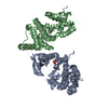


 PDBj
PDBj

 Assembly


 Mass: 65.409 Da / Num. of mol.: 2 / Source method: obtained synthetically / Formula: Zn
 Mass: 96.063 Da / Num. of mol.: 5 / Source method: obtained synthetically / Formula: SO4
 Mass: 35.453 Da / Num. of mol.: 12 / Source method: obtained synthetically / Formula: Cl
 Mass: 62.068 Da / Num. of mol.: 10 / Source method: obtained synthetically / Formula: C2H6O2
 Sample preparation
 / Beamline: BL11-1 / Wavelength: 0.97871, 0.91837
 Processing