 Movie
Movie Controller

[English] 日本語
 Yorodumi
- PDB-1jaz: Crystal Structure of Monoclinic Form of D90E Mutant of Escherichi... -
+ Open data

- Basic information
| Entry | Database: PDB / ID: 1jaz | ||||||
|---|---|---|---|---|---|---|---|
| Title | Crystal Structure of Monoclinic Form of D90E Mutant of Escherichia coli Asparaginase II | ||||||
 Components Components | L-ASPARAGINASE II | ||||||
 Keywords Keywords | HYDROLASE / L-asparaginase / leukemia / zinc-binding site | ||||||
| Function / homology |  Function and homology information Function and homology informationL-asparagine catabolic process / asparaginase / asparaginase activity / outer membrane-bounded periplasmic space / protein homotetramerization / periplasmic space / protein-containing complex / identical protein binding Similarity search - Function | ||||||
| Biological species |  | ||||||
| Method |  X-RAY DIFFRACTION / SYNCHROTRON / MOLECULAR REPLACEMENT / Resolution: 2.27 Å X-RAY DIFFRACTION / SYNCHROTRON / MOLECULAR REPLACEMENT / Resolution: 2.27 Å | ||||||
 Authors Authors | Borek, D. / Kozak, M. / Jaskolski, M. | ||||||
 Citation Citation | Journal: Febs J. / Year: 2014 Title: Crystal structure of active site mutant of antileukemic L-asparaginase reveals conserved zinc-binding site. Authors: Borek, D. / Kozak, M. / Pei, J. / Jaskolski, M. #1: Journal: Proc.Natl.Acad.Sci.USA / Year: 1993Title: Crystal Structure of Escherichia coli L-Asparaginase, An Enzyme Used in Cancer Therapy Authors: Swain, A.L. / Jaskolski, M. / Housset, D. / Rao, J.K. / Wlodawer, A. #2: Journal: FEBS Lett. / Year: 1996Title: A Covalently Bound Catalytic Intermediate in Escherichia coli Asparaginase: Crystal Structure of a Thr-89-Val Mutant Authors: Palm, G.J. / Lubkowski, J. / Derst, C. / Schleper, S. / Rohm, K.H. / Wlodawer, A. #3: Journal: ACTA CRYSTALLOGR.,SECT.D / Year: 2001Title: Structures of Two Highly Homologous Bacterial L-Asparaginases: a Case of Enantiomorphic Space Groups Authors: Jaskolski, M. / Kozak, M. / Lubkowski, J. / Palm, G. / Wlodawer, A. #4: Journal: ACTA BIOCHIM.POL. / Year: 1997Title: Why a "Benign" Mutation Kills Enzyme Activity. Structure-based Analysis of the A176V Mutant of Saccharomyces cerevisiae L-Asparaginase I Authors: Bonthron, D.T. / Jaskolski, M. #5: Journal: BIOCHIM.BIOPHYS.ACTA / Year: 2000Title: Dynamics of a mobile loop at the active site of Escherichia coli Asparaginase Authors: Aung, H.P. / Bocola, M. / Schleper, S. / Rohm, K.H. | ||||||
| History |
| ||||||
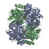
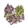
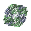
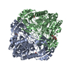
| Remark 300 | BIOMOLECULE: 1 THIS ENTRY CONTAINS THE CRYSTALLOGRAPHIC ASYMMETRIC UNIT WHICH CONSISTS OF 2 CHAIN(S) ...BIOMOLECULE: 1 THIS ENTRY CONTAINS THE CRYSTALLOGRAPHIC ASYMMETRIC UNIT WHICH CONSISTS OF 2 CHAIN(S). SEE REMARK 350 FOR INFORMATION ON GENERATING THE BIOLOGICAL MOLECULE(S). THE BIOLOGICALLY SIGNIFICANT OLIGOMER IS A HOMOTETRAMER WITH 222 SYMMETRY. THE ASYMMETRIC UNIT CONSIST OF MONOMERS A AND B. THE ACTIVE-SITE-COMPETENT DIMERS (A/A' AND B/B') ARE CREATED THROUGH CRYSTALLOGRAPHIC TWO-FOLD ROTATION. |
- Structure visualization
| Structure viewer | Molecule: MolmilJmol/JSmol |
|---|
- Downloads & links
-Download
| PDBx/mmCIF format | 1jaz.cif.gz | 127.7 KB | Display | PDBx/mmCIF format |
|---|---|---|---|---|
| PDB format | pdb1jaz.ent.gz | 99.6 KB | Display | PDB format |
| PDBx/mmJSON format | 1jaz.json.gz | Tree view | PDBx/mmJSON format | |
| Others |  Other downloads Other downloads |
-Validation report
| Arichive directory | https://data.pdbj.org/pub/pdb/validation_reports/ja/1jazftp://data.pdbj.org/pub/pdb/validation_reports/ja/1jaz | HTTPS FTP |
|---|
-Related structure data
| Related structure data |  1ihdC 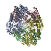 1jjaC  3ecaS C: citing same article ( S: Starting model for refinement |
|---|---|
| Similar structure data |


-Links
 PDBj
PDBj- Assembly
| Deposited unit | 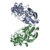
| ||||||||
|---|---|---|---|---|---|---|---|---|---|
| 1 | 
| ||||||||
| Unit cell |
| ||||||||
| Components on special symmetry positions |
| ||||||||
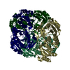
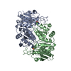
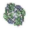
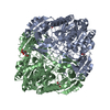
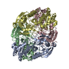
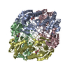
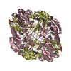
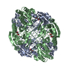
| Details | The biological assembly is a homotetramer. The asymmetric unit contains two asparaginase monomers. The complete tetramer is generated by crystallographic two-fold rotation which relates the two subunits necessary for the creation of the active site. |
-Components
| #1: Protein | Mass: 34640.836 Da / Num. of mol.: 2 / Mutation: D90E Source method: isolated from a genetically manipulated source Source: (gene. exp.) #2: Chemical |   Mass: 65.409 Da / Num. of mol.: 3 / Source method: obtained synthetically / Formula: Zn Mass: 65.409 Da / Num. of mol.: 3 / Source method: obtained synthetically / Formula: Zn#3: Water | ChemComp-HOH / |  Mass: 18.015 Da / Num. of mol.: 136 / Source method: isolated from a natural source / Formula: H2O Mass: 18.015 Da / Num. of mol.: 136 / Source method: isolated from a natural source / Formula: H2OHas protein modification | Y | |
|---|
-Experimental details
-Experiment
| Experiment | Method: X-RAY DIFFRACTION / Number of used crystals: 2 |
|---|
- Sample preparation
| Crystal | Density Matthews: 2.1 Å3/Da / Density % sol: 40.5 % |
|---|---|
| Crystal grow | Temperature: 293 K / Method: vapor diffusion, hanging drop / pH: 6.5 Details: PEG MME 550, MES, zinc sulfate, pH 6.5, 292 K, VAPOR DIFFUSION, HANGING DROP, temperature 293K |
-Data collection
| Diffraction | Mean temperature: 100 K |
|---|---|
| Diffraction source | Source: SYNCHROTRON / Site: MAX II  / Beamline: I711 / Wavelength: 1.104 Å / Beamline: I711 / Wavelength: 1.104 Å |
| Detector | Type: MARRESEARCH / Detector: IMAGE PLATE / Date: Mar 21, 1999 / Details: Vertically focusing cylindrical pre-mirror |
| Radiation | Monochromator: Single asymmetrically cut Si(111) crystal with horizontal diffraction plane. The crystal is bendable for horizontal focusing. Protocol: SINGLE WAVELENGTH / Monochromatic (M) / Laue (L): M / Scattering type: x-ray |
| Radiation wavelength | Wavelength: 1.104 Å / Relative weight: 1 |
| Reflection | Resolution: 2.27→25 Å / Num. all: 26057 / Num. obs: 26057 / % possible obs: 98.6 % / Observed criterion σ(F): 0 / Observed criterion σ(I): -3 / Redundancy: 4.9 % / Biso Wilson estimate: 36.4 Å2 / Rmerge(I) obs: 0.098 / Net I/σ(I): 28.9 |
| Reflection shell | Resolution: 2.27→2.35 Å / Redundancy: 2.9 % / Rmerge(I) obs: 0.143 / Mean I/σ(I) obs: 6.2 / % possible all: 87.3 |
- Processing
| Software |
| ||||||||||||||||||||||||||||||||||||||||||||||||||||||||||||||||||||||||||||||||||||
|---|---|---|---|---|---|---|---|---|---|---|---|---|---|---|---|---|---|---|---|---|---|---|---|---|---|---|---|---|---|---|---|---|---|---|---|---|---|---|---|---|---|---|---|---|---|---|---|---|---|---|---|---|---|---|---|---|---|---|---|---|---|---|---|---|---|---|---|---|---|---|---|---|---|---|---|---|---|---|---|---|---|---|---|---|---|
| Refinement | Method to determine structure: MOLECULAR REPLACEMENT Starting model: MONOMER A FROM NATIVE L-ASPARAGINASE II STRUCTURE - PDB CODE: 3ECA Resolution: 2.27→10 Å / SU B: 11.03497 / SU ML: 0.27657 / Cross valid method: THROUGHOUT / σ(F): 0 / σ(I): -3 / ESU R: 0.38927 / ESU R Free: 0.23674 / Stereochemistry target values: ENGH AND HUBER Details: Maximum likelihood method and TLS parameters were used. Residues 15-35 of monomer A and residues 15-37 of monomer B are not included in the model because of poor electron density and ...Details: Maximum likelihood method and TLS parameters were used. Residues 15-35 of monomer A and residues 15-37 of monomer B are not included in the model because of poor electron density and possible disorder.EACH SUBUNIT (A AND B) COORDINATES ONE ZINC ION. ANOTHER ZINC CATION (AT HALF OCCUPANCY) IS LOCATED CLOSE TO THE CRYSTALLOGRAPHIC TWO-FOLD AXIS AND IS COORDINATED BY TWO COPIES OF SUBUNIT B.
| ||||||||||||||||||||||||||||||||||||||||||||||||||||||||||||||||||||||||||||||||||||
| Displacement parameters | Biso mean: 22.56 Å2
| ||||||||||||||||||||||||||||||||||||||||||||||||||||||||||||||||||||||||||||||||||||
| Refinement step | Cycle: LAST / Resolution: 2.27→10 Å
| ||||||||||||||||||||||||||||||||||||||||||||||||||||||||||||||||||||||||||||||||||||
| Refine LS restraints |
|
