 Movie
Movie Controller

[English] 日本語
 Yorodumi
- PDB-1ho3: CRYSTAL STRUCTURE ANALYSIS OF E. COLI L-ASPARAGINASE II (Y25F MUTANT) -
+ Open data

- Basic information
| Entry | Database: PDB / ID: 1ho3 | ||||||
|---|---|---|---|---|---|---|---|
| Title | CRYSTAL STRUCTURE ANALYSIS OF E. COLI L-ASPARAGINASE II (Y25F MUTANT) | ||||||
 Components Components | L-ASPARAGINASE II | ||||||
 Keywords Keywords | HYDROLASE / asparaginase / leukemia | ||||||
| Function / homology |  Function and homology information Function and homology informationL-asparagine catabolic process / asparaginase / asparaginase activity / outer membrane-bounded periplasmic space / protein homotetramerization / periplasmic space / protein-containing complex / identical protein binding Similarity search - Function | ||||||
| Biological species |  | ||||||
| Method |  X-RAY DIFFRACTION / SYNCHROTRON / MOLECULAR REPLACEMENT / Resolution: 2.5 Å X-RAY DIFFRACTION / SYNCHROTRON / MOLECULAR REPLACEMENT / Resolution: 2.5 Å | ||||||
 Authors Authors | Jaskolski, M. / Kozak, M. / Lubkowski, P. / Palm, J.G. / Wlodawer, A. | ||||||
 Citation Citation | Journal: Acta Crystallogr.,Sect.D / Year: 2001 Title: Structures of two highly homologous bacterial L-asparaginases: a case of enantiomorphic space groups. Authors: Jaskolski, M. / Kozak, M. / Lubkowski, J. / Palm, G. / Wlodawer, A. #1: Journal: ACTA BIOCHIM.POL. / Year: 2000Title: Preliminary Crystallographic Studies of Y25F Mutant of Periplasmic Escherichia coli L-asparaginase Authors: Kozak, M. / Jaskolski, M. / Roehm, K.H. #2: Journal: Proc.Natl.Acad.Sci.USA / Year: 1993Title: Crystal Structure of Escherichia coli L-asparaginase, an Enzyme Used in Cancer Therapy Authors: Swain, A.L. / Jaskolski, M. / Housset, D. / Rao, J.K.M. / Wlodawer, A. | ||||||
| History |
|
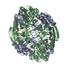
- Structure visualization
| Structure viewer | Molecule: MolmilJmol/JSmol |
|---|
- Downloads & links
-Download
| PDBx/mmCIF format | 1ho3.cif.gz | 132.6 KB | Display | PDBx/mmCIF format |
|---|---|---|---|---|
| PDB format | pdb1ho3.ent.gz | 104.9 KB | Display | PDB format |
| PDBx/mmJSON format | 1ho3.json.gz | Tree view | PDBx/mmJSON format | |
| Others |  Other downloads Other downloads |
-Validation report
| Arichive directory | https://data.pdbj.org/pub/pdb/validation_reports/ho/1ho3ftp://data.pdbj.org/pub/pdb/validation_reports/ho/1ho3 | HTTPS FTP |
|---|
-Related structure data
| Related structure data | 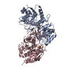 1hfjC  1hfkC  3ecaS S: Starting model for refinement C: citing same article ( |
|---|---|
| Similar structure data |


-Links
 PDBj
PDBj
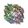
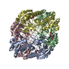
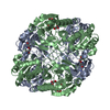
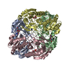
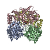
- Assembly
| Deposited unit | 
| ||||||||
|---|---|---|---|---|---|---|---|---|---|
| 1 | 
| ||||||||
| Unit cell |
| ||||||||
| Components on special symmetry positions |
| ||||||||
| Details | The biological assembly is a homotetramer generated from the dimer in the asymmetric unit by the action of crystallographic twofold axis [1, -1,0] |
-Components
| #1: Protein | Mass: 34610.809 Da / Num. of mol.: 2 / Mutation: Y25F Source method: isolated from a genetically manipulated source Source: (gene. exp.) #2: Chemical |   Type: L-peptide linking / Mass: 133.103 Da / Num. of mol.: 2 / Source method: obtained synthetically / Formula: C4H7NO4 Type: L-peptide linking / Mass: 133.103 Da / Num. of mol.: 2 / Source method: obtained synthetically / Formula: C4H7NO4#3: Water | ChemComp-HOH / |  Mass: 18.015 Da / Num. of mol.: 109 / Source method: isolated from a natural source / Formula: H2O Mass: 18.015 Da / Num. of mol.: 109 / Source method: isolated from a natural source / Formula: H2OHas protein modification | Y | |
|---|
-Experimental details
-Experiment
| Experiment | Method: X-RAY DIFFRACTION / Number of used crystals: 1 |
|---|
- Sample preparation
| Crystal | Density Matthews: 2.33 Å3/Da / Density % sol: 47.26 % | ||||||||||||||||||||||||||||||||||||
|---|---|---|---|---|---|---|---|---|---|---|---|---|---|---|---|---|---|---|---|---|---|---|---|---|---|---|---|---|---|---|---|---|---|---|---|---|---|
| Crystal grow | Temperature: 298 K / Method: vapor diffusion, hanging drop / pH: 4.8 Details: MPD, calcium chloride, sodium citrate, pH 4.8, VAPOR DIFFUSION, HANGING DROP, temperature 298K | ||||||||||||||||||||||||||||||||||||
| Crystal | *PLUS Density % sol: 58 % | ||||||||||||||||||||||||||||||||||||
| Crystal grow | *PLUS Temperature: 20 ℃ / Method: vapor diffusion / Details: Kozak, M., (2000) Acta Biochim. Pol., 47, 807. | ||||||||||||||||||||||||||||||||||||
| Components of the solutions | *PLUS
|
-Data collection
| Diffraction | Mean temperature: 290 K |
|---|---|
| Diffraction source | Source: SYNCHROTRON / Site: EMBL/DESY, HAMBURG  / Beamline: X11 / Wavelength: 0.98 Å / Beamline: X11 / Wavelength: 0.98 Å |
| Detector | Type: MARRESEARCH / Detector: IMAGE PLATE / Date: Dec 10, 1995 |
| Radiation | Monochromator: Triangular monochromator bent mirror / Protocol: SINGLE WAVELENGTH / Monochromatic (M) / Laue (L): M / Scattering type: x-ray |
| Radiation wavelength | Wavelength: 0.98 Å / Relative weight: 1 |
| Reflection | Resolution: 2.45→20 Å / Num. all: 74462 / Num. obs: 23517 / % possible obs: 92 % / Observed criterion σ(F): 0 / Observed criterion σ(I): 0 / Redundancy: 3.17 % / Biso Wilson estimate: 15.3 Å2 / Rmerge(I) obs: 0.097 / Net I/σ(I): 7.9 |
| Reflection shell | Resolution: 2.45→2.54 Å / Redundancy: 1.9 % / Rmerge(I) obs: 0.364 / Mean I/σ(I) obs: 2 / % possible all: 85.4 |
| Reflection | *PLUS Num. measured all: 74462 |
| Reflection shell | *PLUS % possible obs: 87 % |
- Processing
| Software |
| ||||||||||||||||||||||||||||||||||||||||
|---|---|---|---|---|---|---|---|---|---|---|---|---|---|---|---|---|---|---|---|---|---|---|---|---|---|---|---|---|---|---|---|---|---|---|---|---|---|---|---|---|---|
| Refinement | Method to determine structure: MOLECULAR REPLACEMENT Starting model: PDB ENTRY 3ECA Resolution: 2.5→10 Å / Rfactor Rfree error: 0.008 / Data cutoff high rms absF: 69969.35 / Cross valid method: THROUGHOUT / σ(F): 2 / σ(I): 1 / Stereochemistry target values: Engh & Huber
| ||||||||||||||||||||||||||||||||||||||||
| Solvent computation | Solvent model: flat model / Bsol: 67.3721 Å2 / ksol: 0.340528 e/Å3 | ||||||||||||||||||||||||||||||||||||||||
| Displacement parameters | Biso mean: 47.4 Å2
| ||||||||||||||||||||||||||||||||||||||||
| Refine analyze |
| ||||||||||||||||||||||||||||||||||||||||
| Refinement step | Cycle: LAST / Resolution: 2.5→10 Å
| ||||||||||||||||||||||||||||||||||||||||
| Refine LS restraints |
| ||||||||||||||||||||||||||||||||||||||||
| LS refinement shell | Resolution: 2.5→2.65 Å / Rfactor Rfree error: 0.037 / Total num. of bins used: 6
| ||||||||||||||||||||||||||||||||||||||||
| Software | *PLUS Name: CNS / Classification: refinement | ||||||||||||||||||||||||||||||||||||||||
| Refinement | *PLUS Highest resolution: 2.5 Å / Lowest resolution: 10 Å / σ(F): 2 / % reflection Rfree: 4.9 % / Rfactor obs: 0.182 | ||||||||||||||||||||||||||||||||||||||||
| Solvent computation | *PLUS | ||||||||||||||||||||||||||||||||||||||||
| Displacement parameters | *PLUS Biso mean: 47.4 Å2 | ||||||||||||||||||||||||||||||||||||||||
| Refine LS restraints | *PLUS
| ||||||||||||||||||||||||||||||||||||||||
| LS refinement shell | *PLUS Rfactor Rfree: 0.355 / % reflection Rfree: 4.6 % / Rfactor Rwork: 0.322 |
