 Movie
Movie Controller

[English] 日本語
 Yorodumi
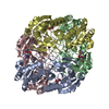
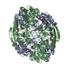
- PDB-3eca: CRYSTAL STRUCTURE OF ESCHERICHIA COLI L-ASPARAGINASE, AN ENZYME U... -
+ Open data

- Basic information
| Entry | Database: PDB / ID: 3eca | |||||||||
|---|---|---|---|---|---|---|---|---|---|---|
| Title | CRYSTAL STRUCTURE OF ESCHERICHIA COLI L-ASPARAGINASE, AN ENZYME USED IN CANCER THERAPY (ELSPAR) | |||||||||
 Components Components | L-asparaginase 2 | |||||||||
 Keywords Keywords | HYDROLASE | |||||||||
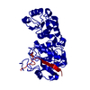
| Function / homology |  Function and homology information Function and homology informationL-asparagine catabolic process / : / asparaginase / asparaginase activity / outer membrane-bounded periplasmic space / protein homotetramerization / periplasmic space / protein-containing complex / identical protein binding Similarity search - Function | |||||||||
| Biological species |  | |||||||||
| Method |  X-RAY DIFFRACTION / MIR / Resolution: 2.4 Å X-RAY DIFFRACTION / MIR / Resolution: 2.4 Å | |||||||||
 Authors Authors | Swain, A.L. / Jaskolski, M. / Housset, D. / Rao, J.K.M. / Wlodawer, A. | |||||||||
 Citation Citation | Journal: Proc.Natl.Acad.Sci.USA / Year: 1993 Title: Crystal structure of Escherichia coli L-asparaginase, an enzyme used in cancer therapy. Authors: Swain, A.L. / Jaskolski, M. / Housset, D. / Rao, J.K. / Wlodawer, A. #1: Journal: J.Biol.Chem. / Year: 1988 Title: Preliminary Crystal Structure of Acinetobacter Glutaminasificans Glutaminase-Asparaginase Authors: Ammon, H.L. / Weber, I.T. / Wlodawer, A. / Harrison, R.W. / Gilliland, G.L. / Murphy, K.C. / Sjolin, L. / Roberts, J. | |||||||||
| History |
|
- Structure visualization
| Structure viewer | Molecule: MolmilJmol/JSmol |
|---|
- Downloads & links
-Download
| PDBx/mmCIF format | 3eca.cif.gz | 256.9 KB | Display | PDBx/mmCIF format |
|---|---|---|---|---|
| PDB format | pdb3eca.ent.gz | 206.8 KB | Display | PDB format |
| PDBx/mmJSON format | 3eca.json.gz | Tree view | PDBx/mmJSON format | |
| Others |  Other downloads Other downloads |
-Validation report
| Arichive directory | https://data.pdbj.org/pub/pdb/validation_reports/ec/3ecaftp://data.pdbj.org/pub/pdb/validation_reports/ec/3eca | HTTPS FTP |
|---|
-Related structure data
| Similar structure data |
|---|


-Links
 PDBj
PDBj
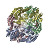
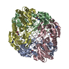
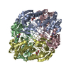
- Assembly
| Deposited unit | 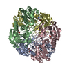
| ||||||||
|---|---|---|---|---|---|---|---|---|---|
| 1 |
| ||||||||
| Unit cell |
|
-Components
| #1: Protein | Mass: 34626.766 Da / Num. of mol.: 4 / Source method: isolated from a natural source Details: The structure was determined for the clinical drug Elspar. The amino acid sequence differed in four places from the sequence of the K12 strain of E. coli asparaginase (V27A, N64D, S252T, T263N). Source: (natural) References: UniProt: A0A377K0N3, UniProt: P00805*PLUS, asparaginase #2: Chemical | ChemComp-ASP /   Type: L-peptide linking / Mass: 133.103 Da / Num. of mol.: 4 / Source method: obtained synthetically / Formula: C4H7NO4 Type: L-peptide linking / Mass: 133.103 Da / Num. of mol.: 4 / Source method: obtained synthetically / Formula: C4H7NO4#3: Water | ChemComp-HOH / |  Mass: 18.015 Da / Num. of mol.: 465 / Source method: isolated from a natural source / Formula: H2O Mass: 18.015 Da / Num. of mol.: 465 / Source method: isolated from a natural source / Formula: H2OHas ligand of interest | N | Has protein modification | Y | |
|---|
-Experimental details
-Experiment
| Experiment | Method: X-RAY DIFFRACTION |
|---|
- Sample preparation
| Crystal | Density Matthews: 2.94 Å3/Da / Density % sol: 58.13 % | |||||||||||||||||||||||||
|---|---|---|---|---|---|---|---|---|---|---|---|---|---|---|---|---|---|---|---|---|---|---|---|---|---|---|
| Crystal grow | Temperature: 293 K / Method: vapor diffusion, hanging drop / pH: 5 / Details: 0.1 M sodium acetate, MPD, PEG3350 | |||||||||||||||||||||||||
| Crystal grow | *PLUS pH: 5 / Method: vapor diffusion, hanging drop | |||||||||||||||||||||||||
| Components of the solutions | *PLUS
|
-Data collection
| Diffraction | Mean temperature: 293 K / Serial crystal experiment: N |
|---|---|
| Diffraction source | Source: ROTATING ANODE / Type: ENRAF-NONIUS FR590 / Wavelength: 1.542 Å |
| Detector | Type: SIEMENS-XENTRONICS / Detector: AREA DETECTOR / Date: Jan 1, 1992 |
| Radiation | Protocol: SINGLE WAVELENGTH / Monochromatic (M) / Laue (L): M / Scattering type: x-ray |
| Radiation wavelength | Wavelength: 1.542 Å / Relative weight: 1 |
| Reflection | Resolution: 2.4→10 Å / % possible obs: 85.3 % / Redundancy: 5.4 % / Rmerge(I) obs: 0.055 |
| Reflection | *PLUS Highest resolution: 2.3 Å / % possible obs: 74 % / Rmerge(I) obs: 0.055 |
| Reflection shell | *PLUS Redundancy: 5.4 % |
- Processing
| Software |
| ||||||||||||||||||||||||||||||||||||||||||||||||||||||||||||
|---|---|---|---|---|---|---|---|---|---|---|---|---|---|---|---|---|---|---|---|---|---|---|---|---|---|---|---|---|---|---|---|---|---|---|---|---|---|---|---|---|---|---|---|---|---|---|---|---|---|---|---|---|---|---|---|---|---|---|---|---|---|
| Refinement | Method to determine structure: MIR / Resolution: 2.4→9.99 Å / Cor.coef. Fo:Fc: 0.974 / SU B: 3.053 / SU ML: 0.072 / Cross valid method: THROUGHOUT / σ(F): 0 / ESU R: 0.252 / Stereochemistry target values: MAXIMUM LIKELIHOOD Details: HYDROGENS ADDED IN RIDING POSITIONS U VALUES: REFINED INDIVIDUALLY
| ||||||||||||||||||||||||||||||||||||||||||||||||||||||||||||
| Solvent computation | Ion probe radii: 0.8 Å / Shrinkage radii: 0.8 Å / VDW probe radii: 1.2 Å / Solvent model: MASK | ||||||||||||||||||||||||||||||||||||||||||||||||||||||||||||
| Displacement parameters | Biso max: 169.82 Å2 / Biso mean: 25.809 Å2 / Biso min: 2.86 Å2
| ||||||||||||||||||||||||||||||||||||||||||||||||||||||||||||
| Refinement step | Cycle: final / Resolution: 2.4→9.99 Å
| ||||||||||||||||||||||||||||||||||||||||||||||||||||||||||||
| Refine LS restraints |
| ||||||||||||||||||||||||||||||||||||||||||||||||||||||||||||
| LS refinement shell | Resolution: 2.4→2.458 Å / Rfactor Rfree error: 0 / Total num. of bins used: 20
| ||||||||||||||||||||||||||||||||||||||||||||||||||||||||||||
| Software | *PLUS Name: X-PLOR / Classification: refinement | ||||||||||||||||||||||||||||||||||||||||||||||||||||||||||||
| Refinement | *PLUS Highest resolution: 2.4 Å / Rfactor obs: 0.149 / Num. reflection obs: 56390 | ||||||||||||||||||||||||||||||||||||||||||||||||||||||||||||
| Solvent computation | *PLUS | ||||||||||||||||||||||||||||||||||||||||||||||||||||||||||||
| Displacement parameters | *PLUS | ||||||||||||||||||||||||||||||||||||||||||||||||||||||||||||
| Refine LS restraints | *PLUS Type: x_angle_d / Dev ideal: 0.054 |
