 Movie
Movie Controller


+ Open data

- Basic information
| Entry | Database: PDB / ID: 4pga | ||||||
|---|---|---|---|---|---|---|---|
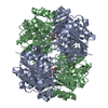
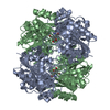
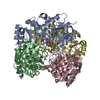
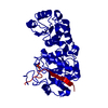
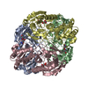
| Title | GLUTAMINASE-ASPARAGINASE FROM PSEUDOMONAS 7A | ||||||
 Components Components | GLUTAMINASE-ASPARAGINASE | ||||||
 Keywords Keywords | BACTERIAL AMIDOHYDROLASE | ||||||
| Function / homology |  Function and homology information Function and homology informationglutamin-(asparagin-)ase / glutamin-(asparagin-)ase activity / : / asparaginase activity / glutaminase activity / periplasmic space Similarity search - Function | ||||||
| Biological species |  Pseudomonas sp. 7A (bacteria) Pseudomonas sp. 7A (bacteria) | ||||||
| Method |  X-RAY DIFFRACTION / MOLECULAR REPLACEMENT / Resolution: 1.7 Å X-RAY DIFFRACTION / MOLECULAR REPLACEMENT / Resolution: 1.7 Å | ||||||
 Authors Authors | Jakob, C.G. / Lewinski, K. / Lacount, M.W. / Roberts, J. / Lebioda, L. | ||||||
 Citation Citation | Journal: Biochemistry / Year: 1997 Title: Ion binding induces closed conformation in Pseudomonas 7A glutaminase-asparaginase (PGA): crystal structure of the PGA-SO4(2-)-NH4+ complex at 1.7 A resolution. Authors: Jakob, C.G. / Lewinski, K. / LaCount, M.W. / Roberts, J. / Lebioda, L. #1: Journal: To be PublishedTitle: Refined Crystal Structure of Acinetobacter Glutaminasificans Glutaminase-Asparaginase Authors: Lubkowski, J. / Wlodawer, A. / Housset, D. / Weber, I.T. / Ammon, H.L. / Murphy, K.C. / Swain, A.L. #2: Journal: Proc.Natl.Acad.Sci.USA / Year: 1993Title: Crystal Structure of Escherichia Coli L-Asparaginase, an Enzyme Used in Cancer Therapy Authors: Swain, A.L. / Jaskolski, M. / Housset, D. / Rao, J.K. / Wlodawer, A. #3: Journal: FEBS Lett. / Year: 1993Title: A Left-Handed Crossover Involved in Amidohydrolase Catalysis. Crystal Structure of Erwinia Chrysanthemi L-Asparaginase with Bound L-Aspartate Authors: Miller, M. / Rao, J.K. / Wlodawer, A. / Gribskov, M.R. | ||||||
| History |
|
- Structure visualization
| Structure viewer | Molecule: MolmilJmol/JSmol |
|---|
- Downloads & links
-Download
| PDBx/mmCIF format | 4pga.cif.gz | 141.9 KB | Display | PDBx/mmCIF format |
|---|---|---|---|---|
| PDB format | pdb4pga.ent.gz | 112.1 KB | Display | PDB format |
| PDBx/mmJSON format | 4pga.json.gz | Tree view | PDBx/mmJSON format | |
| Others |  Other downloads Other downloads |
-Validation report
| Arichive directory | https://data.pdbj.org/pub/pdb/validation_reports/pg/4pgaftp://data.pdbj.org/pub/pdb/validation_reports/pg/4pga | HTTPS FTP |
|---|
-Related structure data
| Related structure data |  3ecaS S: Starting model for refinement |
|---|---|
| Similar structure data |

-Links
 PDBj
PDBj
- Assembly
| Deposited unit | 
| ||||||||
|---|---|---|---|---|---|---|---|---|---|
| 1 | 
| ||||||||
| Unit cell |
|
-Components
| #1: Protein | Mass: 36252.918 Da / Num. of mol.: 2 Source method: isolated from a genetically manipulated source Details: AMIDOHYDROLASE, ASPARAGINASE / Source: (gene. exp.) Pseudomonas sp. 7A (bacteria) / References: UniProt: P10182, glutamin-(asparagin-)ase#2: Chemical |   Mass: 96.063 Da / Num. of mol.: 2 / Source method: obtained synthetically / Formula: SO4 Mass: 96.063 Da / Num. of mol.: 2 / Source method: obtained synthetically / Formula: SO4#3: Chemical |   Mass: 18.038 Da / Num. of mol.: 2 / Source method: obtained synthetically / Formula: H4N Mass: 18.038 Da / Num. of mol.: 2 / Source method: obtained synthetically / Formula: H4N#4: Water | ChemComp-HOH / |  Mass: 18.015 Da / Num. of mol.: 405 / Source method: isolated from a natural source / Formula: H2O Mass: 18.015 Da / Num. of mol.: 405 / Source method: isolated from a natural source / Formula: H2O |
|---|
-Experimental details
-Experiment
| Experiment | Method: X-RAY DIFFRACTION / Number of used crystals: 1 |
|---|
- Sample preparation
| Crystal | Density Matthews: 2.6 Å3/Da / Density % sol: 51.52 % |
|---|---|
| Crystal grow | pH: 4.6 / Details: pH 4.6 |
| Crystal grow | *PLUS Method: vapor diffusion, hanging drop |
| Components of the solutions | *PLUS Conc.: 2.0 M / Common name: ammonium sulfate |
-Data collection
| Diffraction | Mean temperature: 293 K |
|---|---|
| Diffraction source | Source: ROTATING ANODE / Type: RIGAKU RUH2R / Wavelength: 1.5418 |
| Detector | Type: RIGAKU / Detector: IMAGE PLATE / Date: Nov 1, 1995 / Details: MIRRORS |
| Radiation | Monochromator: NI FILTER / Monochromatic (M) / Laue (L): M / Scattering type: x-ray |
| Radiation wavelength | Wavelength: 1.5418 Å / Relative weight: 1 |
| Reflection | Resolution: 1.71→39.31 Å / Num. obs: 68971 / % possible obs: 86 % / Observed criterion σ(I): 1 / Redundancy: 2.6 % / Rmerge(I) obs: 0.075 / Rsym value: 0.075 / Net I/σ(I): 19.9 |
| Reflection shell | Resolution: 1.71→1.77 Å / Redundancy: 1.6 % / Rmerge(I) obs: 0.261 / Mean I/σ(I) obs: 2 / Rsym value: 0.261 / % possible all: 62 |
| Reflection | *PLUS Num. all: 79799 / Num. measured all: 178566 |
| Reflection shell | *PLUS % possible obs: 62 % |
- Processing
| Software |
| ||||||||||||||||||||||||||||||||||||||||||||||||||||||||||||
|---|---|---|---|---|---|---|---|---|---|---|---|---|---|---|---|---|---|---|---|---|---|---|---|---|---|---|---|---|---|---|---|---|---|---|---|---|---|---|---|---|---|---|---|---|---|---|---|---|---|---|---|---|---|---|---|---|---|---|---|---|---|
| Refinement | Method to determine structure: MOLECULAR REPLACEMENT Starting model: PDB ENTRY 3ECA Resolution: 1.7→10 Å / Data cutoff high absF: 10000000 / Data cutoff low absF: 0.001 / Cross valid method: THROUGHOUT / σ(F): 2
| ||||||||||||||||||||||||||||||||||||||||||||||||||||||||||||
| Refinement step | Cycle: LAST / Resolution: 1.7→10 Å
| ||||||||||||||||||||||||||||||||||||||||||||||||||||||||||||
| Refine LS restraints |
| ||||||||||||||||||||||||||||||||||||||||||||||||||||||||||||
| LS refinement shell | Resolution: 1.71→10 Å / Total num. of bins used: 1
| ||||||||||||||||||||||||||||||||||||||||||||||||||||||||||||
| Xplor file |
| ||||||||||||||||||||||||||||||||||||||||||||||||||||||||||||
| Software | *PLUS Name: X-PLOR / Classification: refinement | ||||||||||||||||||||||||||||||||||||||||||||||||||||||||||||
| Refinement | *PLUS | ||||||||||||||||||||||||||||||||||||||||||||||||||||||||||||
| Solvent computation | *PLUS | ||||||||||||||||||||||||||||||||||||||||||||||||||||||||||||
| Displacement parameters | *PLUS | ||||||||||||||||||||||||||||||||||||||||||||||||||||||||||||
| Refine LS restraints | *PLUS
| ||||||||||||||||||||||||||||||||||||||||||||||||||||||||||||
| LS refinement shell | *PLUS Rfactor obs: 0.199 |
