 Movie
Movie Controller

[English] 日本語
 Yorodumi
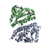
- PDB-3pga: STRUCTURAL CHARACTERIZATION OF PSEUDOMONAS 7A GLUTAMINASE-ASPARAGINASE -
+ Open data

- Basic information
| Entry | Database: PDB / ID: 3pga | ||||||
|---|---|---|---|---|---|---|---|
| Title | STRUCTURAL CHARACTERIZATION OF PSEUDOMONAS 7A GLUTAMINASE-ASPARAGINASE | ||||||
 Components Components | GLUTAMINASE-ASPARAGINASE | ||||||
 Keywords Keywords | BACTERIAL AMIDOHYDROLASE | ||||||
| Function / homology |  Function and homology information Function and homology informationglutamin-(asparagin-)ase / glutamin-(asparagin-)ase activity / : / asparaginase activity / glutaminase activity / periplasmic space Similarity search - Function | ||||||
| Biological species |  Pseudomonas sp. 7A (bacteria) Pseudomonas sp. 7A (bacteria) | ||||||
| Method |  X-RAY DIFFRACTION / Resolution: 2 Å X-RAY DIFFRACTION / Resolution: 2 Å | ||||||
 Authors Authors | Lubkowski, J. / Wlodawer, A. / Ammon, H.L. / Copeland, T.D. / Swain, A.L. | ||||||
 Citation Citation | Journal: Biochemistry / Year: 1994 Title: Structural characterization of Pseudomonas 7A glutaminase-asparaginase. Authors: Lubkowski, J. / Wlodawer, A. / Ammon, H.L. / Copeland, T.D. / Swain, A.L. #1: Journal: To be PublishedTitle: Refined Crystal Structure of Acinetobacter Glutaminasificans Glutaminase-Asparaginase Authors: Lubkowski, J. / Wlodawer, A. / Housset, D. / Weber, I.T. / Ammon, H.L. / Murphy, K.C. / Swain, A.L. #2: Journal: FEBS Lett. / Year: 1993Title: A Left-Handed Crossover Involved in Amidohydrolase Catalysis: Crystal Structure of Erwinia Chrysanthemi L-Asparaginase with Bound L-Aspartate Authors: Miller, M. / Rao, J.K.M. / Wlodawer, A. / Gribskov, M.R. #3: Journal: Proc.Natl.Acad.Sci.USA / Year: 1993Title: Crystal Structure of E. Coli L-Asparaginase, an Enzyme Used in Cancer Therapy Authors: Swain, A.L. / Jaskolski, M. / Housset, D. / Rao, J.K.M. / Wlodawer, A. #4: Journal: Acta Crystallogr.,Sect.B / Year: 1983Title: The Molecular Symmetry of Glutaminase-Asparaginases: Rotation Function Studies of the Pseudomonas 7A and Acinetobacter Enzymes Authors: Ammon, H.L. / Murphy, K.C. / Sjolin, L. / Wlodawer, A. / Holcenberg, J.S. / Roberts, J. #5: Journal: Biochemistry / Year: 1978Title: Amino Acid Sequence of the Diazooxonorleucine Binding Site of Acinetobacter and Pseudomonas 7A Glutaminase-Asparaginase Enzymes Authors: Holcenberg, J.S. / Ericsson, L. / Roberts, J. #6: Journal: J.Mol.Biol. / Year: 1977Title: Characterization of Crystals of L-Glutaminase-Asparaginase from Acinetobacter Glutaminasificans and Pseudomonas 7A Authors: Wlodawer, A. / Roberts, J. / Holcenberg, J.S. | ||||||
| History |
|
- Structure visualization
| Structure viewer | Molecule: MolmilJmol/JSmol |
|---|
- Downloads & links
-Download
| PDBx/mmCIF format | 3pga.cif.gz | 275.2 KB | Display | PDBx/mmCIF format |
|---|---|---|---|---|
| PDB format | pdb3pga.ent.gz | 216 KB | Display | PDB format |
| PDBx/mmJSON format | 3pga.json.gz | Tree view | PDBx/mmJSON format | |
| Others |  Other downloads Other downloads |
-Validation report
| Arichive directory | https://data.pdbj.org/pub/pdb/validation_reports/pg/3pgaftp://data.pdbj.org/pub/pdb/validation_reports/pg/3pga | HTTPS FTP |
|---|
-Related structure data
| Similar structure data |
|---|


-Links
 PDBj
PDBj- Assembly
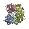
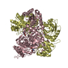
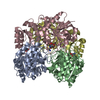
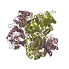
| Deposited unit | 
| ||||||||
|---|---|---|---|---|---|---|---|---|---|
| 1 |
| ||||||||
| Unit cell |
| ||||||||
| Atom site foot note | 1: GLY 1 19 - THR 1 20C OMEGA = 140.34 PEPTIDE BOND DEVIATES SIGNIFICANTLY FROM TRANS CONFORMATION |
-Components
| #1: Protein | Mass: 35978.660 Da / Num. of mol.: 4 Source method: isolated from a genetically manipulated source Source: (gene. exp.) Pseudomonas sp. 7A (bacteria) / References: UniProt: P10182, asparaginase#2: Water | ChemComp-HOH / |  Mass: 18.015 Da / Num. of mol.: 418 / Source method: isolated from a natural source / Formula: H2O Mass: 18.015 Da / Num. of mol.: 418 / Source method: isolated from a natural source / Formula: H2OSequence details | THE SEQUENCE HAS NOT BEEN REPORTED. IT WAS DERIVED FROM THE SEQUENCE OF LUBKOWSKI ET AL., 1994, ...THE SEQUENCE HAS NOT BEEN REPORTED. IT WAS DERIVED FROM THE SEQUENCE OF LUBKOWSKI ET AL., 1994, LISTED IN THE JRNL ARTICLE ABOVE. | |
|---|
-Experimental details
-Experiment
| Experiment | Method: X-RAY DIFFRACTION |
|---|
- Sample preparation
| Crystal | Density Matthews: 2.28 Å3/Da / Density % sol: 46.17 % | |||||||||||||||||||||||||
|---|---|---|---|---|---|---|---|---|---|---|---|---|---|---|---|---|---|---|---|---|---|---|---|---|---|---|
| Crystal grow | *PLUS Temperature: 4 ℃ / pH: 7.2 / Method: vapor diffusion, sitting drop | |||||||||||||||||||||||||
| Components of the solutions | *PLUS
|
-Data collection
| Reflection | Num. obs: 81878 / % possible obs: 86 % / Observed criterion σ(I): 1.5 |
|---|---|
| Reflection | *PLUS Highest resolution: 1.8 Å / Lowest resolution: 15 Å / Num. measured all: 212153 / Rmerge(I) obs: 0.073 |
- Processing
| Software |
| ||||||||||||||||||||||||||||||||||||||||||||||||||||||||||||||||||||||||||||||||
|---|---|---|---|---|---|---|---|---|---|---|---|---|---|---|---|---|---|---|---|---|---|---|---|---|---|---|---|---|---|---|---|---|---|---|---|---|---|---|---|---|---|---|---|---|---|---|---|---|---|---|---|---|---|---|---|---|---|---|---|---|---|---|---|---|---|---|---|---|---|---|---|---|---|---|---|---|---|---|---|---|---|
| Refinement | Resolution: 2→10 Å / σ(F): 2.5 Details: IN THIS PDB COORDINATE ENTRY, RESIDUES 1 - 7 IN ALL FOUR CHAINS ARE OMITTED DUE TO DISORDER. FOR RESIDUES 20 - 39, TWO ALTERNATE CONFORMATIONS ARE INCLUDED. (CLOSED CONFORMATION = ALTERNATE ...Details: IN THIS PDB COORDINATE ENTRY, RESIDUES 1 - 7 IN ALL FOUR CHAINS ARE OMITTED DUE TO DISORDER. FOR RESIDUES 20 - 39, TWO ALTERNATE CONFORMATIONS ARE INCLUDED. (CLOSED CONFORMATION = ALTERNATE CONFORMATION CODE C; OPEN CONFORMATION = ALTERNATE CONFORMATION CODE O). THE X, Y, Z COORDINATES AND B FACTORS FOR RESIDUES 20 - 39 OF CHAIN 2 IN THE OPEN CONFORMATION WERE REFINED AGAINST X-RAY DATA. THE COORDINATES FOR THE CLOSED CONFORMATION OF RESIDUES 20 - 39 OF CHAIN 2 AND BOTH CONFORMATIONS OF RESIDUES 20 - 39 IN CHAINS 1, 3, AND 4 WERE NOT REFINED BUT RATHER MODELLED; THE ATOMS OF THESE RESIDUES HAVE AN OCCUPANCY OF 0.0 AND B FACTOR OF 20.0. NOTE THAT RESIDUE 28 WAS TREATED AS GLY (RATHER THAN ALA) IN THE CLOSED CONFORMATION IN THE COORDINATES THAT WERE DEPOSITED. IT HAS BEEN IDENTIFIED AS ALA IN THIS ENTRY. BECAUSE OF THIS THERE ARE NO COORDINATES FOR ATOM CB OF ALA 28 IN THE CLOSED CONFORMATION.
| ||||||||||||||||||||||||||||||||||||||||||||||||||||||||||||||||||||||||||||||||
| Refinement step | Cycle: LAST / Resolution: 2→10 Å
| ||||||||||||||||||||||||||||||||||||||||||||||||||||||||||||||||||||||||||||||||
| Refine LS restraints |
| ||||||||||||||||||||||||||||||||||||||||||||||||||||||||||||||||||||||||||||||||
| Software | *PLUS Name: X-PLOR/PROFFT / Classification: refinement | ||||||||||||||||||||||||||||||||||||||||||||||||||||||||||||||||||||||||||||||||
| Refinement | *PLUS Rfactor obs: 0.165 | ||||||||||||||||||||||||||||||||||||||||||||||||||||||||||||||||||||||||||||||||
| Solvent computation | *PLUS | ||||||||||||||||||||||||||||||||||||||||||||||||||||||||||||||||||||||||||||||||
| Displacement parameters | *PLUS Biso mean: 30.74 Å2 | ||||||||||||||||||||||||||||||||||||||||||||||||||||||||||||||||||||||||||||||||
| Refine LS restraints | *PLUS
|
