 Movie
Movie Controller

[English] 日本語
 Yorodumi
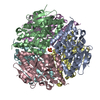
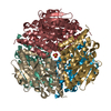
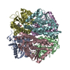
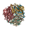
- PDB-2j5s: Structural of ABDH, a beta-diketone hydrolase from the Cyanobacte... -
+ Open data

- Basic information
| Entry | Database: PDB / ID: 2j5s | ||||||
|---|---|---|---|---|---|---|---|
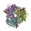
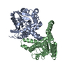
| Title | Structural of ABDH, a beta-diketone hydrolase from the Cyanobacterium Anabaena sp. PCC 7120 bound to (S)-3-oxocyclohexyl acetic acid | ||||||
 Components Components | BETA-DIKETONE HYDROLASE | ||||||
 Keywords Keywords | HYDROLASE / ENZYME EVOLUTION / C-C BOND HYDROLASE / LYASE / CROTONASE / BIOCATALYSIS / BETA-DIKETONE | ||||||
| Function / homology |  Function and homology information Function and homology informationEnoyl-CoA hydratase/isomerase / Enoyl-CoA hydratase/isomerase / 2-enoyl-CoA Hydratase; Chain A, domain 1 / 2-enoyl-CoA Hydratase; Chain A, domain 1 / ClpP/crotonase-like domain superfamily / Alpha-Beta Complex / Alpha Beta Similarity search - Domain/homology | ||||||
| Biological species |  ANABAENA SP. (bacteria) ANABAENA SP. (bacteria) | ||||||
| Method |  X-RAY DIFFRACTION / SYNCHROTRON / MOLECULAR REPLACEMENT / Resolution: 1.57 Å X-RAY DIFFRACTION / SYNCHROTRON / MOLECULAR REPLACEMENT / Resolution: 1.57 Å | ||||||
 Authors Authors | Bennett, J.P. / Whittingham, J.L. / Brzozowski, A.M. / Leonard, P.M. / Grogan, G. | ||||||
 Citation Citation | Journal: Biochemistry / Year: 2007 Title: Structural Characterisation of a Beta Diketone Hydrolase from the Cyanobacterium Anabaena Sp. Pcc 7120 in Native and Product Bound Forms, a Coenzyme A-Independent Member of the Crotonase Suprafamily Authors: Bennett, J.P. / Whittingham, J.L. / Brzozowski, A.M. / Leonard, P.M. / Grogan, G. | ||||||
| History |
|
- Structure visualization
| Structure viewer | Molecule: MolmilJmol/JSmol |
|---|
- Downloads & links
-Download
| PDBx/mmCIF format | 2j5s.cif.gz | 127.9 KB | Display | PDBx/mmCIF format |
|---|---|---|---|---|
| PDB format | pdb2j5s.ent.gz | 98.5 KB | Display | PDB format |
| PDBx/mmJSON format | 2j5s.json.gz | Tree view | PDBx/mmJSON format | |
| Others |  Other downloads Other downloads |
-Validation report
| Arichive directory | https://data.pdbj.org/pub/pdb/validation_reports/j5/2j5sftp://data.pdbj.org/pub/pdb/validation_reports/j5/2j5s | HTTPS FTP |
|---|
-Related structure data
| Related structure data |  2j5gC  1o8uS  2j5j C: citing same article ( S: Starting model for refinement |
|---|---|
| Similar structure data |


-Links
 PDBj
PDBj- Assembly
| Deposited unit | 
| ||||||||||||||||||||||||||||||
|---|---|---|---|---|---|---|---|---|---|---|---|---|---|---|---|---|---|---|---|---|---|---|---|---|---|---|---|---|---|---|---|
| 1 | 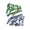
| ||||||||||||||||||||||||||||||
| Unit cell |
| ||||||||||||||||||||||||||||||
| Components on special symmetry positions |
|
-Components
| #1: Protein | Mass: 30067.037 Da / Num. of mol.: 2 Source method: isolated from a genetically manipulated source Details: PROTEIN WAS CO-CRYSTALLISED WITH SUBSTRATE / Source: (gene. exp.) ANABAENA SP. (bacteria) / Strain: PCC 7120 / Production host: #2: Chemical | 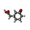  Mass: 156.179 Da / Num. of mol.: 2 / Source method: obtained synthetically / Formula: C8H12O3 Mass: 156.179 Da / Num. of mol.: 2 / Source method: obtained synthetically / Formula: C8H12O3#3: Chemical |   Mass: 58.693 Da / Num. of mol.: 2 / Source method: obtained synthetically / Formula: Ni Mass: 58.693 Da / Num. of mol.: 2 / Source method: obtained synthetically / Formula: Ni#4: Water | ChemComp-HOH / |  Mass: 18.015 Da / Num. of mol.: 490 / Source method: isolated from a natural source / Formula: H2O Mass: 18.015 Da / Num. of mol.: 490 / Source method: isolated from a natural source / Formula: H2OSequence details | SEQUENCE CONTAINS N-TERMINAL CLEAVABLE HIS TAG OF SIX HISTIDINE RESIDUES. | |
|---|
-Experimental details
-Experiment
| Experiment | Method: X-RAY DIFFRACTION / Number of used crystals: 1 |
|---|
- Sample preparation
| Crystal | Density Matthews: 2.04 Å3/Da / Density % sol: 39.61 % |
|---|---|
| Crystal grow | pH: 7.5 Details: 0.1 M BIS-TRIS PROPANE PH 7.5, 20% PEG 3350, 0.2 M SODIUM MALONATE, 0.01 M BICYCLO[2.2.2]OCTANE-2,6-DIONE |
-Data collection
| Diffraction | Mean temperature: 100 K |
|---|---|
| Diffraction source | Source: SYNCHROTRON / Site: ESRF  / Beamline: ID23-1 / Wavelength: 1.07 / Beamline: ID23-1 / Wavelength: 1.07 |
| Detector | Type: ADSC CCD / Detector: CCD / Date: May 20, 2006 / Details: TOROIDAL MIRROR |
| Radiation | Monochromator: SILICON (1 1 1) CHANNEL- CUT / Protocol: SINGLE WAVELENGTH / Monochromatic (M) / Laue (L): M / Scattering type: x-ray |
| Radiation wavelength | Wavelength: 1.07 Å / Relative weight: 1 |
| Reflection | Resolution: 1.57→40.19 Å / Num. obs: 64435 / % possible obs: 98.9 % / Redundancy: 10.1 % / Rmerge(I) obs: 0.06 / Net I/σ(I): 32.04 |
| Reflection shell | Resolution: 1.57→1.63 Å / Redundancy: 4.8 % / Rmerge(I) obs: 0.27 / Mean I/σ(I) obs: 2.93 / % possible all: 89.9 |
- Processing
| Software |
| ||||||||||||||||||||||||||||||||||||||||||||||||||||||||||||||||||||||||||||||||||||||||||||||||||||||||||||||||||||||||||||||||||||||||||||||||||||||||||||||||||||||||||||||||||||||
|---|---|---|---|---|---|---|---|---|---|---|---|---|---|---|---|---|---|---|---|---|---|---|---|---|---|---|---|---|---|---|---|---|---|---|---|---|---|---|---|---|---|---|---|---|---|---|---|---|---|---|---|---|---|---|---|---|---|---|---|---|---|---|---|---|---|---|---|---|---|---|---|---|---|---|---|---|---|---|---|---|---|---|---|---|---|---|---|---|---|---|---|---|---|---|---|---|---|---|---|---|---|---|---|---|---|---|---|---|---|---|---|---|---|---|---|---|---|---|---|---|---|---|---|---|---|---|---|---|---|---|---|---|---|---|---|---|---|---|---|---|---|---|---|---|---|---|---|---|---|---|---|---|---|---|---|---|---|---|---|---|---|---|---|---|---|---|---|---|---|---|---|---|---|---|---|---|---|---|---|---|---|---|---|
| Refinement | Method to determine structure: MOLECULAR REPLACEMENT Starting model: PDB ENTRY 1O8U Resolution: 1.57→40.19 Å / Cor.coef. Fo:Fc: 0.975 / Cor.coef. Fo:Fc free: 0.962 / SU B: 1.227 / SU ML: 0.045 / Cross valid method: THROUGHOUT / ESU R: 0.074 / ESU R Free: 0.076 / Stereochemistry target values: MAXIMUM LIKELIHOOD / Details: HYDROGENS HAVE BEEN ADDED IN THE RIDING POSITIONS.
| ||||||||||||||||||||||||||||||||||||||||||||||||||||||||||||||||||||||||||||||||||||||||||||||||||||||||||||||||||||||||||||||||||||||||||||||||||||||||||||||||||||||||||||||||||||||
| Solvent computation | Ion probe radii: 0.8 Å / Shrinkage radii: 0.8 Å / VDW probe radii: 1.4 Å / Solvent model: MASK | ||||||||||||||||||||||||||||||||||||||||||||||||||||||||||||||||||||||||||||||||||||||||||||||||||||||||||||||||||||||||||||||||||||||||||||||||||||||||||||||||||||||||||||||||||||||
| Displacement parameters | Biso mean: 14.19 Å2
| ||||||||||||||||||||||||||||||||||||||||||||||||||||||||||||||||||||||||||||||||||||||||||||||||||||||||||||||||||||||||||||||||||||||||||||||||||||||||||||||||||||||||||||||||||||||
| Refinement step | Cycle: LAST / Resolution: 1.57→40.19 Å
| ||||||||||||||||||||||||||||||||||||||||||||||||||||||||||||||||||||||||||||||||||||||||||||||||||||||||||||||||||||||||||||||||||||||||||||||||||||||||||||||||||||||||||||||||||||||
| Refine LS restraints |
|