 Movie
Movie Controller

[English] 日本語
 Yorodumi
- PDB-1szo: Crystal Structure Analysis of the 6-Oxo Camphor Hydrolase His122A... -
+ Open data

- Basic information
| Entry | Database: PDB / ID: 1szo | ||||||
|---|---|---|---|---|---|---|---|
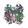
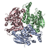
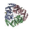
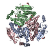
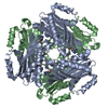
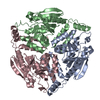
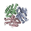
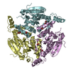
| Title | Crystal Structure Analysis of the 6-Oxo Camphor Hydrolase His122Ala Mutant Bound to Its Natural Product (2S,4S)-alpha-Campholinic Acid | ||||||
 Components Components | 6-oxocamphor hydrolase | ||||||
 Keywords Keywords | HYDROLASE / Enzyme-Product Complex | ||||||
| Function / homology |  Function and homology information Function and homology information6-oxocamphor hydrolase / hydrolase activity, acting on carbon-carbon bonds, in ketonic substances Similarity search - Function | ||||||
| Biological species |  Rhodococcus sp. NCIMB 9784 (bacteria) Rhodococcus sp. NCIMB 9784 (bacteria) | ||||||
| Method |  X-RAY DIFFRACTION / SYNCHROTRON / MOLECULAR REPLACEMENT / Resolution: 1.9 Å X-RAY DIFFRACTION / SYNCHROTRON / MOLECULAR REPLACEMENT / Resolution: 1.9 Å | ||||||
 Authors Authors | Leonard, P.M. / Grogan, G. | ||||||
 Citation Citation | Journal: J.Biol.Chem. / Year: 2004 Title: Structure of 6-oxo camphor hydrolase H122A mutant bound to its natural product, (2S,4S)-alpha-campholinic acid: mutant structure suggests an atypical mode of transition state binding for a crotonase homolog. Authors: Leonard, P.M. / Grogan, G. #1: Journal: J.Biol.Chem. / Year: 2003Title: The 2- Crystal Structure of 6-Oxo Camphor Hydrolase. NEW STRUCTURAL DIVERSITY IN THE CROTONASE SUPERFAMILY. Authors: Whittingham, J.L. / Turkenburg, J.P. / Verma, C.S. / Walsh, M.A. / Grogan, G. #2: Journal: J.Biol.Chem. / Year: 2001Title: The Desymmetrization of Bicyclic beta-Diketones by an Enzymatic Retro-Claisen Reaction. A NEW REACTION OF THE CROTONASE SUPERFAMILY. Authors: Grogan, G. / Roberts, G.A. / Bougioukou, D. / Turner, N.J. / Flitsch, S.L. #3: Journal: Angew.Chem.Int.Ed.Engl. / Year: 2001Title: An Asymmetric Enzyme-Catalysed Retro-Claisen Reaction for the Desymmetrisation of Cyclic beta-Diketones. Authors: Grogan, G. / Graf, J. / Jones, A. / Parsons, S. / Turner, N.J. / Flitsch, S.L. | ||||||
| History |
|
- Structure visualization
| Structure viewer | Molecule: MolmilJmol/JSmol |
|---|
- Downloads & links
-Download
| PDBx/mmCIF format | 1szo.cif.gz | 610 KB | Display | PDBx/mmCIF format |
|---|---|---|---|---|
| PDB format | pdb1szo.ent.gz | 500.9 KB | Display | PDB format |
| PDBx/mmJSON format | 1szo.json.gz | Tree view | PDBx/mmJSON format | |
| Others |  Other downloads Other downloads |
-Validation report
| Arichive directory | https://data.pdbj.org/pub/pdb/validation_reports/sz/1szoftp://data.pdbj.org/pub/pdb/validation_reports/sz/1szo | HTTPS FTP |
|---|
-Related structure data
| Related structure data |  1o8uS S: Starting model for refinement |
|---|---|
| Similar structure data |


-Links
 PDBj
PDBj- Assembly
| Deposited unit | 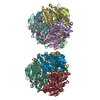
| ||||||||
|---|---|---|---|---|---|---|---|---|---|
| 1 | 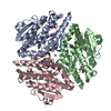
| ||||||||
| 2 | 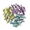
| ||||||||
| 3 | 
| ||||||||
| 4 | 
| ||||||||
| 5 | 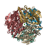
| ||||||||
| 6 | 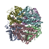
| ||||||||
| Unit cell |
| ||||||||
| Details | The biological assembly is a trimer, of which there are four complete copies in the asymmetric unit. |
-Components
| #1: Protein | Mass: 28446.172 Da / Num. of mol.: 12 / Mutation: H122A Source method: isolated from a genetically manipulated source Source: (gene. exp.) Rhodococcus sp. NCIMB 9784 (bacteria) / Gene: camK / Plasmid: pET-26b (Novagen) / Species (production host): Escherichia coli / Production host: #2: Chemical | ChemComp-CAX / (   Mass: 186.248 Da / Num. of mol.: 12 / Source method: obtained synthetically / Formula: C10H18O3 Mass: 186.248 Da / Num. of mol.: 12 / Source method: obtained synthetically / Formula: C10H18O3#3: Chemical | ChemComp-CA /   Mass: 40.078 Da / Num. of mol.: 4 / Source method: obtained synthetically / Formula: Ca Mass: 40.078 Da / Num. of mol.: 4 / Source method: obtained synthetically / Formula: Ca#4: Water | ChemComp-HOH / |  Mass: 18.015 Da / Num. of mol.: 1929 / Source method: isolated from a natural source / Formula: H2O Mass: 18.015 Da / Num. of mol.: 1929 / Source method: isolated from a natural source / Formula: H2O |
|---|
-Experimental details
-Experiment
| Experiment | Method: X-RAY DIFFRACTION / Number of used crystals: 1 |
|---|
- Sample preparation
| Crystal | Density Matthews: 2.2 Å3/Da / Density % sol: 42.9 % |
|---|---|
| Crystal grow | Temperature: 290 K / Method: vapor diffusion, hanging drop / pH: 5.6 Details: PEG MME 2000, calcium acetate, MES, pH 5.6, VAPOR DIFFUSION, HANGING DROP, temperature 290K |
-Data collection
| Diffraction | Mean temperature: 100 K |
|---|---|
| Diffraction source | Source: SYNCHROTRON / Site: ESRF  / Beamline: ID14-3 / Wavelength: 0.931 Å / Beamline: ID14-3 / Wavelength: 0.931 Å |
| Detector | Type: MARRESEARCH / Detector: CCD / Date: Nov 15, 2003 / Details: Toroidal mirror |
| Radiation | Monochromator: Diamond (111), Ge(220) / Protocol: SINGLE WAVELENGTH / Monochromatic (M) / Laue (L): M / Scattering type: x-ray |
| Radiation wavelength | Wavelength: 0.931 Å / Relative weight: 1 |
| Reflection | Resolution: 1.9→30 Å / Num. all: 229538 / Num. obs: 229538 / % possible obs: 100 % / Observed criterion σ(F): 2 / Observed criterion σ(I): 2 / Redundancy: 3.8 % / Biso Wilson estimate: 21.2 Å2 / Rmerge(I) obs: 0.06 / Rsym value: 0.047 / Net I/σ(I): 29.2 |
| Reflection shell | Resolution: 1.9→1.96 Å / Redundancy: 3.7 % / Rmerge(I) obs: 0.282 / Mean I/σ(I) obs: 4.9 / Num. unique all: 19032 / Rsym value: 0.241 / % possible all: 100 |
- Processing
| Software |
| |||||||||||||||||||||||||
|---|---|---|---|---|---|---|---|---|---|---|---|---|---|---|---|---|---|---|---|---|---|---|---|---|---|---|
| Refinement | Method to determine structure: MOLECULAR REPLACEMENT Starting model: PDB Entry 1O8U Resolution: 1.9→30 Å / Isotropic thermal model: Isotropic / Cross valid method: THROUGHOUT / σ(F): 0 / σ(I): 0 / Stereochemistry target values: Engh & Huber
| |||||||||||||||||||||||||
| Displacement parameters | Biso mean: 18.7 Å2
| |||||||||||||||||||||||||
| Refine analyze |
| |||||||||||||||||||||||||
| Refinement step | Cycle: LAST / Resolution: 1.9→30 Å
| |||||||||||||||||||||||||
| Refine LS restraints |
| |||||||||||||||||||||||||
| LS refinement shell | Resolution: 1.9→1.947 Å
|
