| Entry | Database: PDB / ID: 4u18
|
|---|
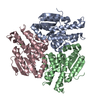
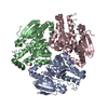
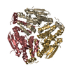
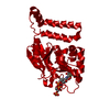
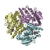
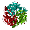
| Title | Crystal structure of human peroxisomal delta3,delta2, enoyl-CoA isomerase (ISO-ECI2) |
|---|
 Components Components | Enoyl-CoA delta isomerase 2, mitochondrial |
|---|
 Keywords Keywords | ISOMERASE / PECI / enoy-CoA isomerase / crotonase / beta-oxidation |
|---|
| Function / homology |  Function and homology information Function and homology information
Beta-oxidation of very long chain fatty acids / Delta3-Delta2-enoyl-CoA isomerase / delta(3)-delta(2)-enoyl-CoA isomerase activity / fatty-acyl-CoA binding / fatty acid catabolic process / fatty acid beta-oxidation / peroxisomal matrix / Peroxisomal protein import / peroxisome / mitochondrion ...Beta-oxidation of very long chain fatty acids / Delta3-Delta2-enoyl-CoA isomerase / delta(3)-delta(2)-enoyl-CoA isomerase activity / fatty-acyl-CoA binding / fatty acid catabolic process / fatty acid beta-oxidation / peroxisomal matrix / Peroxisomal protein import / peroxisome / mitochondrion / membrane / cytosolSimilarity search - Function Acyl-CoA-binding protein, ACBP, conserved site / Acyl-CoA-binding (ACB) domain signature. / Acyl-CoA-binding protein, ACBP / Acyl-CoA binding protein superfamily / : / Acyl CoA binding protein / Acyl-CoA-binding (ACB) domain profile. / Lyase 2-enoyl-coa Hydratase, Chain A, domain 2 / Lyase 2-enoyl-coa Hydratase; Chain A, domain 2 / Enoyl-CoA hydratase, C-terminal ...Acyl-CoA-binding protein, ACBP, conserved site / Acyl-CoA-binding (ACB) domain signature. / Acyl-CoA-binding protein, ACBP / Acyl-CoA binding protein superfamily / : / Acyl CoA binding protein / Acyl-CoA-binding (ACB) domain profile. / Lyase 2-enoyl-coa Hydratase, Chain A, domain 2 / Lyase 2-enoyl-coa Hydratase; Chain A, domain 2 / Enoyl-CoA hydratase, C-terminal / Enoyl-CoA hydratase/isomerase / Enoyl-CoA hydratase/isomerase / 2-enoyl-CoA Hydratase; Chain A, domain 1 / 2-enoyl-CoA Hydratase; Chain A, domain 1 / FERM/acyl-CoA-binding protein superfamily / ClpP/crotonase-like domain superfamily / Alpha-Beta Complex / Orthogonal Bundle / Mainly Alpha / Alpha BetaSimilarity search - Domain/homology |
|---|
| Biological species |  Homo sapiens (human) Homo sapiens (human) |
|---|
| Method |  X-RAY DIFFRACTION / SYNCHROTRON / MOLECULAR REPLACEMENT / Resolution: 2.64 Å X-RAY DIFFRACTION / SYNCHROTRON / MOLECULAR REPLACEMENT / Resolution: 2.64 Å |
|---|
 Authors Authors | Onwukwe, G.U. / Koski, M.K. / Wierenga, R.K. |
|---|
| Funding support |  Germany, 1items Germany, 1items | Organization | Grant number | Country |
|---|
| Biostruct-X | N283570 | Germany |
|
|---|
 Citation Citation | Journal: Febs J. / Year: 2015
Title: Human Delta (3) , Delta (2) -enoyl-CoA isomerase, type 2: a structural enzymology study on the catalytic role of its ACBP domain and helix-10.
Authors: Onwukwe, G.U. / Kursula, P. / Koski, M.K. / Schmitz, W. / Wierenga, R.K. |
|---|
| History | | Deposition | Jul 15, 2014 | Deposition site: RCSB / Processing site: PDBE |
|---|
| Revision 1.0 | Dec 24, 2014 | Provider: repository / Type: Initial release |
|---|
| Revision 1.1 | Dec 31, 2014 | Group: Database references |
|---|
| Revision 1.2 | Jan 14, 2015 | Group: Database references |
|---|
| Revision 1.3 | Feb 25, 2015 | Group: Database references |
|---|
| Revision 1.4 | May 8, 2024 | Group: Data collection / Database references / Refinement description
Category: chem_comp_atom / chem_comp_bond ...chem_comp_atom / chem_comp_bond / database_2 / struct_ncs_dom_lim
Item: _database_2.pdbx_DOI / _database_2.pdbx_database_accession ..._database_2.pdbx_DOI / _database_2.pdbx_database_accession / _struct_ncs_dom_lim.beg_auth_comp_id / _struct_ncs_dom_lim.beg_label_asym_id / _struct_ncs_dom_lim.beg_label_comp_id / _struct_ncs_dom_lim.beg_label_seq_id / _struct_ncs_dom_lim.end_auth_comp_id / _struct_ncs_dom_lim.end_label_asym_id / _struct_ncs_dom_lim.end_label_comp_id / _struct_ncs_dom_lim.end_label_seq_id |
|---|
|
|---|
 Movie
Movie Controller

 Yorodumi
 Open data
 Basic information
 Structure visualization
 Downloads & links
 Other downloads


 PDBj
PDBj
 Assembly
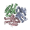


 Mass: 35.453 Da / Num. of mol.: 3 / Source method: isolated from a natural source / Formula: Cl
 Mass: 92.094 Da / Num. of mol.: 1 / Source method: obtained synthetically / Formula: C3H8O3
 Mass: 18.015 Da / Num. of mol.: 21 / Source method: isolated from a natural source / Formula: H2O
 Sample preparation
 / Beamline: ID29 / Wavelength: 0.976 Å
 Processing