 Movie
Movie Controller

[English] 日本語
 Yorodumi
- PDB-6slb: Crystal structure of isomerase PaaG with trans-3,4-didehydroadipyl-CoA -
+ Open data

- Basic information
| Entry | Database: PDB / ID: 6slb | ||||||
|---|---|---|---|---|---|---|---|
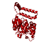
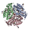
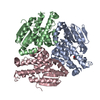
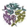
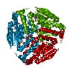
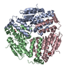
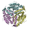
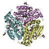
| Title | Crystal structure of isomerase PaaG with trans-3,4-didehydroadipyl-CoA | ||||||
 Components Components | Enoyl-CoA hydratase/carnithine racemase | ||||||
 Keywords Keywords | ISOMERASE / phenylacetic acid catabolism / tropodithietic acid / crotonase | ||||||
| Function / homology | Enoyl-CoA hydratase, C-terminal / Enoyl-CoA hydratase/isomerase / Enoyl-CoA hydratase/isomerase / catalytic activity / ClpP/crotonase-like domain superfamily / Chem-T3D / Enoyl-CoA hydratase/carnithine racemase Function and homology information Function and homology information | ||||||
| Biological species |   Thermus thermophilus JL-18 (bacteria) Thermus thermophilus JL-18 (bacteria) | ||||||
| Method |  X-RAY DIFFRACTION / SYNCHROTRON / MOLECULAR REPLACEMENT / Resolution: 1.88 Å X-RAY DIFFRACTION / SYNCHROTRON / MOLECULAR REPLACEMENT / Resolution: 1.88 Å | ||||||
 Authors Authors | Saleem-Batcha, R. / Spieker, M. / Teufel, R. | ||||||
| Funding support |  Germany, 1items Germany, 1items
| ||||||
 Citation Citation | Journal: Acs Chem.Biol. / Year: 2019 Title: Structural and Mechanistic Basis of an Oxepin-CoA Forming Isomerase in Bacterial Primary and Secondary Metabolism. Authors: Spieker, M. / Saleem-Batcha, R. / Teufel, R. | ||||||
| History |
|
- Structure visualization
| Structure viewer | Molecule: MolmilJmol/JSmol |
|---|
- Downloads & links
-Download
| PDBx/mmCIF format | 6slb.cif.gz | 70.1 KB | Display | PDBx/mmCIF format |
|---|---|---|---|---|
| PDB format | pdb6slb.ent.gz | Display | PDB format | |
| PDBx/mmJSON format | 6slb.json.gz | Tree view | PDBx/mmJSON format | |
| Others |  Other downloads Other downloads |
-Validation report
| Arichive directory | https://data.pdbj.org/pub/pdb/validation_reports/sl/6slbftp://data.pdbj.org/pub/pdb/validation_reports/sl/6slb | HTTPS FTP |
|---|
-Related structure data
| Related structure data |  6sl9C 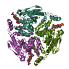 6slaC  3hrxS C: citing same article ( S: Starting model for refinement |
|---|---|
| Similar structure data |


-Links
 PDBj
PDBj- Assembly
| Deposited unit | 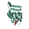
| ||||||||
|---|---|---|---|---|---|---|---|---|---|
| 1 | 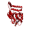
| ||||||||
| Unit cell |
| ||||||||
| Components on special symmetry positions |
|
-Components
| #1: Protein | Mass: 27861.174 Da / Num. of mol.: 1 Source method: isolated from a genetically manipulated source Source: (gene. exp.) Thermus thermophilus JL-18 (bacteria) / Gene: TtJL18_0099 / Production host: |
|---|---|
| #2: Chemical | ChemComp-T3D / (~{  Mass: 893.644 Da / Num. of mol.: 1 / Source method: obtained synthetically / Formula: C27H42N7O19P3S / Feature type: SUBJECT OF INVESTIGATION Mass: 893.644 Da / Num. of mol.: 1 / Source method: obtained synthetically / Formula: C27H42N7O19P3S / Feature type: SUBJECT OF INVESTIGATION |
| #3: Water | ChemComp-HOH /  Mass: 18.015 Da / Num. of mol.: 120 / Source method: isolated from a natural source / Formula: H2O Mass: 18.015 Da / Num. of mol.: 120 / Source method: isolated from a natural source / Formula: H2O |
| Has ligand of interest | Y |
-Experimental details
-Experiment
| Experiment | Method: X-RAY DIFFRACTION / Number of used crystals: 1 |
|---|
- Sample preparation
| Crystal | Density Matthews: 2.04 Å3/Da / Density % sol: 39.7 % |
|---|---|
| Crystal grow | Temperature: 297 K / Method: vapor diffusion, sitting drop Details: 50 mM KH2PO4, pH 4.5, 20-22% PEG 3350, 3% (w/v) NDSB-201 and 20% glycerol (v/v) |
-Data collection
| Diffraction | Mean temperature: 100 K / Serial crystal experiment: N |
|---|---|
| Diffraction source | Source: SYNCHROTRON / Site: SLS  / Beamline: X06SA / Wavelength: 1 Å / Beamline: X06SA / Wavelength: 1 Å |
| Detector | Type: DECTRIS EIGER X 16M / Detector: PIXEL / Date: Jun 30, 2019 |
| Radiation | Protocol: SINGLE WAVELENGTH / Monochromatic (M) / Laue (L): M / Scattering type: x-ray |
| Radiation wavelength | Wavelength: 1 Å / Relative weight: 1 |
| Reflection | Resolution: 1.88→44.01 Å / Num. obs: 18671 / % possible obs: 99.41 % / Redundancy: 2 % / CC1/2: 0.997 / Net I/σ(I): 22.48 |
| Reflection shell | Resolution: 1.88→1.947 Å / Rmerge(I) obs: 0.2998 / Num. unique obs: 1747 |
- Processing
| Software |
| |||||||||||||||||||||||||||||||||||||||||||||||||||||||||||||||||||||||||||||||||||||||||||||||||||||||||||||||||||||||||||||||||||||||||||||||||||||||||||||||||||||
|---|---|---|---|---|---|---|---|---|---|---|---|---|---|---|---|---|---|---|---|---|---|---|---|---|---|---|---|---|---|---|---|---|---|---|---|---|---|---|---|---|---|---|---|---|---|---|---|---|---|---|---|---|---|---|---|---|---|---|---|---|---|---|---|---|---|---|---|---|---|---|---|---|---|---|---|---|---|---|---|---|---|---|---|---|---|---|---|---|---|---|---|---|---|---|---|---|---|---|---|---|---|---|---|---|---|---|---|---|---|---|---|---|---|---|---|---|---|---|---|---|---|---|---|---|---|---|---|---|---|---|---|---|---|---|---|---|---|---|---|---|---|---|---|---|---|---|---|---|---|---|---|---|---|---|---|---|---|---|---|---|---|---|---|---|---|---|
| Refinement | Method to determine structure: MOLECULAR REPLACEMENT Starting model: 3HRX Resolution: 1.88→44.01 Å / Cor.coef. Fo:Fc: 0.97 / Cor.coef. Fo:Fc free: 0.955 / SU B: 3.185 / SU ML: 0.095 / Cross valid method: THROUGHOUT / ESU R: 0.156 / ESU R Free: 0.145 Details: Hydrogens have been added in their riding positions
| |||||||||||||||||||||||||||||||||||||||||||||||||||||||||||||||||||||||||||||||||||||||||||||||||||||||||||||||||||||||||||||||||||||||||||||||||||||||||||||||||||||
| Solvent computation | Ion probe radii: 0.8 Å / Shrinkage radii: 0.8 Å / VDW probe radii: 1.2 Å | |||||||||||||||||||||||||||||||||||||||||||||||||||||||||||||||||||||||||||||||||||||||||||||||||||||||||||||||||||||||||||||||||||||||||||||||||||||||||||||||||||||
| Displacement parameters | Biso mean: 35.196 Å2
| |||||||||||||||||||||||||||||||||||||||||||||||||||||||||||||||||||||||||||||||||||||||||||||||||||||||||||||||||||||||||||||||||||||||||||||||||||||||||||||||||||||
| Refinement step | Cycle: LAST / Resolution: 1.88→44.01 Å
| |||||||||||||||||||||||||||||||||||||||||||||||||||||||||||||||||||||||||||||||||||||||||||||||||||||||||||||||||||||||||||||||||||||||||||||||||||||||||||||||||||||
| Refine LS restraints |
| |||||||||||||||||||||||||||||||||||||||||||||||||||||||||||||||||||||||||||||||||||||||||||||||||||||||||||||||||||||||||||||||||||||||||||||||||||||||||||||||||||||
| LS refinement shell |
|
