ACETATE ION / CITRATE ANION / Enoyl-CoA hydratase/isomerase family protein / Enoyl-CoA hydratase/isomerase family protein Similarity search - Component
#258 - Jun 2021 Glucocorticoid Receptor and Dexamethasone similarity (1)
#89 - May 2007 Aconitase and Iron Regulatory Protein 1 similarity (1)
-
Assembly
Deposited unit
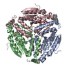
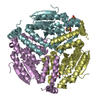
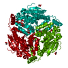
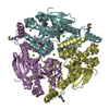
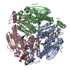
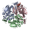
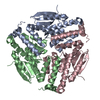
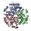
A: Enoyl-CoA hydratase/isomerase family protein B: Enoyl-CoA hydratase/isomerase family protein C: Enoyl-CoA hydratase/isomerase family protein D: Enoyl-CoA hydratase/isomerase family protein E: Enoyl-CoA hydratase/isomerase family protein F: Enoyl-CoA hydratase/isomerase family protein hetero molecules
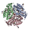
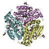
A: Enoyl-CoA hydratase/isomerase family protein B: Enoyl-CoA hydratase/isomerase family protein C: Enoyl-CoA hydratase/isomerase family protein hetero molecules
D: Enoyl-CoA hydratase/isomerase family protein E: Enoyl-CoA hydratase/isomerase family protein F: Enoyl-CoA hydratase/isomerase family protein hetero molecules
Resolution: 1.817→30 Å / Num. all: 151590 / Num. obs: 151590 / % possible obs: 97.3 % / Observed criterion σ(I): -3 / Redundancy: 3.9 % / Biso Wilson estimate: 36.1 Å2 / Rmerge(I) obs: 0.076 / Net I/σ(I): 13.9
Reflection shell
Resolution: 1.817→1.85 Å / Redundancy: 3.9 % / Rmerge(I) obs: 0.61 / Mean I/σ(I) obs: 2.2 / Num. unique all: 7431 / % possible all: 95.7
-
Processing
Software
Name
Version
Classification
Blu-Ice
Max
datacollection
PHENIX
modelbuilding
REFMAC
5.5.0109
refinement
HKL-2000
datareduction
HKL-2000
datascaling
PHENIX
phasing
Refinement
Method to determine structure: MOLECULAR REPLACEMENT Starting model: the starting model was the structure solved by SAD in a different space group Resolution: 1.817→29.016 Å / Cor.coef. Fo:Fc: 0.968 / Cor.coef. Fo:Fc free: 0.958 / SU B: 5.135 / SU ML: 0.071 / Isotropic thermal model: Isotropic / Cross valid method: THROUGHOUT / σ(F): 0 / ESU R: 0.113 / ESU R Free: 0.108 / Stereochemistry target values: MAXIMUM LIKELIHOOD / Details: HYDROGENS HAVE BEEN ADDED IN THE RIDING POSITIONS
Rfactor
Num. reflection
% reflection
Selection details
Rfree
0.19353
7600
5 %
RANDOM
Rwork
0.16211
-
-
-
obs
0.16369
143966
96.75 %
-
all
-
143966
-
-
Solvent computation
Ion probe radii: 0.8 Å / Shrinkage radii: 0.8 Å / VDW probe radii: 1.4 Å / Solvent model: MASK
Displacement parameters
Biso mean: 25.282 Å2
Baniso -1
Baniso -2
Baniso -3
1-
2.02 Å2
-0.66 Å2
0.3 Å2
2-
-
-1.48 Å2
1.26 Å2
3-
-
-
-0.25 Å2
Refine analyze
Luzzati coordinate error obs: 0.2 Å
Refinement step
Cycle: LAST / Resolution: 1.817→29.016 Å
Protein
Nucleic acid
Ligand
Solvent
Total
Num. atoms
11686
0
112
930
12728
Refine LS restraints
Refine-ID
Type
Dev ideal
Dev ideal target
Number
X-RAY DIFFRACTION
r_bond_refined_d
0.017
0.022
12071
X-RAY DIFFRACTION
r_bond_other_d
0.001
0.02
8166
X-RAY DIFFRACTION
r_angle_refined_deg
1.559
1.966
16279
X-RAY DIFFRACTION
r_angle_other_deg
0.94
3
19989
X-RAY DIFFRACTION
r_dihedral_angle_1_deg
5.587
5
1551
X-RAY DIFFRACTION
r_dihedral_angle_2_deg
37.113
24.257
498
X-RAY DIFFRACTION
r_dihedral_angle_3_deg
13.839
15
2094
X-RAY DIFFRACTION
r_dihedral_angle_4_deg
19.184
15
66
X-RAY DIFFRACTION
r_chiral_restr
0.092
0.2
1860
X-RAY DIFFRACTION
r_gen_planes_refined
0.007
0.02
13471
X-RAY DIFFRACTION
r_gen_planes_other
0.001
0.02
2381
X-RAY DIFFRACTION
r_nbd_refined
X-RAY DIFFRACTION
r_nbd_other
X-RAY DIFFRACTION
r_nbtor_refined
X-RAY DIFFRACTION
r_nbtor_other
X-RAY DIFFRACTION
r_xyhbond_nbd_refined
X-RAY DIFFRACTION
r_xyhbond_nbd_other
X-RAY DIFFRACTION
r_metal_ion_refined
X-RAY DIFFRACTION
r_metal_ion_other
X-RAY DIFFRACTION
r_symmetry_vdw_refined
X-RAY DIFFRACTION
r_symmetry_vdw_other
X-RAY DIFFRACTION
r_symmetry_hbond_refined
X-RAY DIFFRACTION
r_symmetry_hbond_other
X-RAY DIFFRACTION
r_symmetry_metal_ion_refined
X-RAY DIFFRACTION
r_symmetry_metal_ion_other
X-RAY DIFFRACTION
r_mcbond_it
0.852
1.5
7681
X-RAY DIFFRACTION
r_mcbond_other
0.259
1.5
3186
X-RAY DIFFRACTION
r_mcangle_it
1.567
2
12288
X-RAY DIFFRACTION
r_scbond_it
2.744
3
4390
X-RAY DIFFRACTION
r_scangle_it
4.504
4.5
3991
X-RAY DIFFRACTION
r_rigid_bond_restr
X-RAY DIFFRACTION
r_sphericity_free
X-RAY DIFFRACTION
r_sphericity_bonded
LS refinement shell
Resolution: 1.817→1.864 Å / Total num. of bins used: 20
Rfactor
Num. reflection
% reflection
Rfree
0.291
538
-
Rwork
0.24
9729
-
obs
-
-
88.31 %
Refinement TLS params.
Method: refined / Refine-ID: X-RAY DIFFRACTION
ID
L11 (°2)
L12 (°2)
L13 (°2)
L22 (°2)
L23 (°2)
L33 (°2)
S11 (Å °)
S12 (Å °)
S13 (Å °)
S21 (Å °)
S22 (Å °)
S23 (Å °)
S31 (Å °)
S32 (Å °)
S33 (Å °)
T11 (Å2)
T12 (Å2)
T13 (Å2)
T22 (Å2)
T23 (Å2)
T33 (Å2)
Origin x (Å)
Origin y (Å)
Origin z (Å)
1
0.6123
0.123
-0.2068
1.3343
-0.2105
0.8054
-0.0583
0.0939
-0.0608
-0.1732
0.0406
-0.2653
0.0269
0.0789
0.0177
0.0357
-0.0044
0.0468
0.0696
-0.0191
0.0677
80.6874
73.938
114.5508
2
0.5669
0.0104
-0.395
0.6982
0.3024
0.9803
-0.0233
-0.0754
-0.1133
0.1264
-0.0261
-0.0482
0.1385
-0.0051
0.0494
0.0635
-0.0128
-0.0132
0.046
0.0148
0.0302
63.548
59.9947
52.1702
3
0.9644
0.4597
-0.6504
0.9536
-0.2315
1.0999
0.1601
-0.0582
0.1351
0.1399
-0.0456
0.0058
-0.2484
0.0003
-0.1145
0.0807
-0.0069
0.0258
0.0117
-0.0139
0.0359
69.0803
93.5119
49.6267
4
0.5064
0.1532
0.1285
1.4645
0.2258
0.8065
-0.0796
0.0915
0.0738
-0.2226
0.0467
0.2821
-0.0297
-0.1044
0.0329
0.0609
-0.0051
-0.0674
0.0882
0.0129
0.0779
29.8096
70.0369
69.953
5
0.5943
0.0195
0.3174
0.7692
-0.2964
0.8551
-0.0106
-0.0655
0.1072
0.0863
-0.0237
0.0564
-0.1128
-0.0031
0.0343
0.0435
-0.011
0.0052
0.0494
-0.0264
0.0273
46.7533
83.208
96.7649
6
0.2623
0.3106
0.1582
0.6072
0.0238
0.6345
0.0917
-0.0496
-0.0687
0.084
-0.0221
0.0073
0.2006
0.0026
-0.0696
0.0738
0.0011
-0.0258
0.0512
0.0113
0.0658
41.9726
50.7316
94.0989
Refinement TLS group
ID
Refine-ID
Refine TLS-ID
Auth asym-ID
Auth seq-ID
1
X-RAY DIFFRACTION
1
A
1 - 257
2
X-RAY DIFFRACTION
2
B
1 - 257
3
X-RAY DIFFRACTION
3
C
0 - 257
4
X-RAY DIFFRACTION
4
D
1 - 257
5
X-RAY DIFFRACTION
5
E
1 - 257
6
X-RAY DIFFRACTION
6
F
1 - 267
+
About Yorodumi
-
News
-
Feb 9, 2022. New format data for meta-information of EMDB entries
New format data for meta-information of EMDB entries
Version 3 of the EMDB header file is now the official format.
The previous official version 1.9 will be removed from the archive.
In the structure databanks used in Yorodumi, some data are registered as the other names, "COVID-19 virus" and "2019-nCoV". Here are the details of the virus and the list of structure data.
Jan 31, 2019. EMDB accession codes are about to change! (news from PDBe EMDB page)
EMDB accession codes are about to change! (news from PDBe EMDB page)
The allocation of 4 digits for EMDB accession codes will soon come to an end. Whilst these codes will remain in use, new EMDB accession codes will include an additional digit and will expand incrementally as the available range of codes is exhausted. The current 4-digit format prefixed with “EMD-” (i.e. EMD-XXXX) will advance to a 5-digit format (i.e. EMD-XXXXX), and so on. It is currently estimated that the 4-digit codes will be depleted around Spring 2019, at which point the 5-digit format will come into force.
The EM Navigator/Yorodumi systems omit the EMD- prefix.
Related info.:Q: What is EMD? / ID/Accession-code notation in Yorodumi/EM Navigator
Yorodumi is a browser for structure data from EMDB, PDB, SASBDB, etc.
This page is also the successor to EM Navigator detail page, and also detail information page/front-end page for Omokage search.
The word "yorodu" (or yorozu) is an old Japanese word meaning "ten thousand". "mi" (miru) is to see.
Related info.:EMDB / PDB / SASBDB / Comparison of 3 databanks / Yorodumi Search / Aug 31, 2016. New EM Navigator & Yorodumi / Yorodumi Papers / Jmol/JSmol / Function and homology information / Changes in new EM Navigator and Yorodumi
 Movie
Movie Controller

 Yorodumi
 Open data
 Basic information
 Components
 Keywords
 Function and homology information
 X-RAY DIFFRACTION /
  Authors
 Citation
 Structure visualization
 Downloads & links
 Other downloads
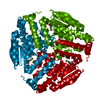

 PDBj
PDBj


 Assembly
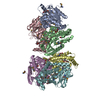

 Mass: 189.100 Da / Num. of mol.: 4 / Source method: obtained synthetically / Formula: C6H5O7
 Mass: 194.226 Da / Num. of mol.: 4 / Source method: obtained synthetically / Formula: C8H18O5 / Comment: precipitant*YM
 Mass: 59.044 Da / Num. of mol.: 2 / Source method: obtained synthetically / Formula: C2H3O2
 Mass: 18.015 Da / Num. of mol.: 930 / Source method: isolated from a natural source / Formula: H2O
 Sample preparation
 / Beamline: 21-ID-D / Wavelength: 1.04006 Å
 Processing