 Movie
Movie Controller

[English] 日本語
 Yorodumi
- PDB-2qin: Stenotrophomonas maltophilia L1 Metallo-beta-Lactamase Asp-120 Cy... -
+ Open data

- Basic information
| Entry | Database: PDB / ID: 2qin | ||||||
|---|---|---|---|---|---|---|---|
| Title | Stenotrophomonas maltophilia L1 Metallo-beta-Lactamase Asp-120 Cys mutant | ||||||
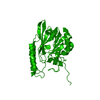
 Components Components | Metallo-beta-lactamase L1 | ||||||
 Keywords Keywords | HYDROLASE / METALLO-BETA-LACTAMASE / BINUCLEAR / DINUCLEAR | ||||||
| Function / homology |  Function and homology information Function and homology informationantibiotic catabolic process / beta-lactamase activity / beta-lactamase / periplasmic space / response to antibiotic / zinc ion binding Similarity search - Function | ||||||
| Biological species |  Stenotrophomonas maltophilia (bacteria) Stenotrophomonas maltophilia (bacteria) | ||||||
| Method |  X-RAY DIFFRACTION / SYNCHROTRON / MOLECULAR REPLACEMENT / Resolution: 1.76 Å X-RAY DIFFRACTION / SYNCHROTRON / MOLECULAR REPLACEMENT / Resolution: 1.76 Å | ||||||
 Authors Authors | Spencer, J. | ||||||
 Citation Citation | Journal: Biochemistry / Year: 2007 Title: Structural Basis for the Role of Asp-120 in Metallo-beta-lactamases. Authors: Crisp, J. / Conners, R. / Garrity, J.D. / Carenbauer, A.L. / Crowder, M.W. / Spencer, J. #1: Journal: J.Biol.Chem. / Year: 2004 Title: Metal Binding Asp-120 in Metallo-beta-lactamase L1 from Stenotrophomonas maltophilia Plays a Crucial Role in Catalysis Authors: Garrity, J.D. / Carenbauer, A.L. / Herron, L.R. / Crowder, M.W. | ||||||
| History |
|
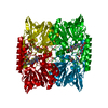
- Structure visualization
| Structure viewer | Molecule: MolmilJmol/JSmol |
|---|
- Downloads & links
-Download
| PDBx/mmCIF format | 2qin.cif.gz | 236.9 KB | Display | PDBx/mmCIF format |
|---|---|---|---|---|
| PDB format | pdb2qin.ent.gz | 185.9 KB | Display | PDB format |
| PDBx/mmJSON format | 2qin.json.gz | Tree view | PDBx/mmJSON format | |
| Others |  Other downloads Other downloads |
-Validation report
| Arichive directory | https://data.pdbj.org/pub/pdb/validation_reports/qi/2qinftp://data.pdbj.org/pub/pdb/validation_reports/qi/2qin | HTTPS FTP |
|---|
-Related structure data
| Related structure data |  2qjsC  1smlS S: Starting model for refinement C: citing same article ( |
|---|---|
| Similar structure data |


-Links
 PDBj
PDBj

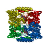
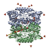
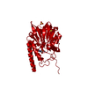
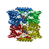
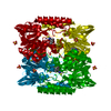
- Assembly
| Deposited unit | 
| ||||||||
|---|---|---|---|---|---|---|---|---|---|
| 1 |
| ||||||||
| 2 | 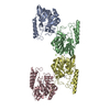
| ||||||||
| 3 | 
| ||||||||
| Unit cell |
|
-Components
| #1: Protein | Mass: 28728.508 Da / Num. of mol.: 4 / Mutation: D120C Source method: isolated from a genetically manipulated source Source: (gene. exp.) Stenotrophomonas maltophilia (bacteria)Strain: IID 1275 / Gene: L1 / Plasmid: pUB5832 / Production host: #2: Chemical | ChemComp-ZN /   Mass: 65.409 Da / Num. of mol.: 9 / Source method: obtained synthetically / Formula: Zn Mass: 65.409 Da / Num. of mol.: 9 / Source method: obtained synthetically / Formula: Zn#3: Chemical |   Mass: 24.305 Da / Num. of mol.: 2 / Source method: obtained synthetically / Formula: Mg Mass: 24.305 Da / Num. of mol.: 2 / Source method: obtained synthetically / Formula: Mg#4: Water | ChemComp-HOH / |  Mass: 18.015 Da / Num. of mol.: 1290 / Source method: isolated from a natural source / Formula: H2O Mass: 18.015 Da / Num. of mol.: 1290 / Source method: isolated from a natural source / Formula: H2OHas protein modification | Y | |
|---|
-Experimental details
-Experiment
| Experiment | Method: X-RAY DIFFRACTION / Number of used crystals: 1 |
|---|
- Sample preparation
| Crystal | Density Matthews: 2.33 Å3/Da / Density % sol: 47.31 % |
|---|---|
| Crystal grow | Temperature: 293 K / Method: vapor diffusion, hanging drop / pH: 7.5 Details: 0.1M Tris.Cl, 0.2M MgCl2, 18% PEG 4000, 5% MPD, pH 7.5, VAPOR DIFFUSION, HANGING DROP, temperature 293K |
-Data collection
| Diffraction | Mean temperature: 100 K |
|---|---|
| Diffraction source | Source: SYNCHROTRON / Site: SRS  / Beamline: PX14.1 / Wavelength: 1.488 Å / Beamline: PX14.1 / Wavelength: 1.488 Å |
| Detector | Type: ADSC QUANTUM 4 / Detector: CCD / Date: Sep 9, 2003 |
| Radiation | Protocol: SINGLE WAVELENGTH / Monochromatic (M) / Laue (L): M / Scattering type: x-ray |
| Radiation wavelength | Wavelength: 1.488 Å / Relative weight: 1 |
| Reflection | Resolution: 1.76→30 Å / Num. all: 104385 / Num. obs: 98730 / % possible obs: 94.6 % / Redundancy: 5.7 % / Biso Wilson estimate: 18.795 Å2 / Rmerge(I) obs: 0.067 / Net I/σ(I): 24 |
| Reflection shell | Resolution: 1.76→1.84 Å / Redundancy: 3.5 % / Rmerge(I) obs: 0.162 / Mean I/σ(I) obs: 7 / Num. unique all: 13052 / % possible all: 69.9 |
- Processing
| Software |
| ||||||||||||||||||||||||||||||||||||||||||||||||||||||||||||||||||||||||||||||||||||||||||||||||||||
|---|---|---|---|---|---|---|---|---|---|---|---|---|---|---|---|---|---|---|---|---|---|---|---|---|---|---|---|---|---|---|---|---|---|---|---|---|---|---|---|---|---|---|---|---|---|---|---|---|---|---|---|---|---|---|---|---|---|---|---|---|---|---|---|---|---|---|---|---|---|---|---|---|---|---|---|---|---|---|---|---|---|---|---|---|---|---|---|---|---|---|---|---|---|---|---|---|---|---|---|---|---|
| Refinement | Method to determine structure: MOLECULAR REPLACEMENT Starting model: PDB ENTRY 1SML Resolution: 1.76→26.84 Å / Cor.coef. Fo:Fc: 0.957 / Cor.coef. Fo:Fc free: 0.938 / SU B: 2.187 / SU ML: 0.072 / Cross valid method: THROUGHOUT / σ(F): 0 / ESU R: 0.128 / ESU R Free: 0.122 / Stereochemistry target values: MAXIMUM LIKELIHOOD / Details: HYDROGENS HAVE BEEN ADDED IN THE RIDING POSITIONS
| ||||||||||||||||||||||||||||||||||||||||||||||||||||||||||||||||||||||||||||||||||||||||||||||||||||
| Solvent computation | Ion probe radii: 0.8 Å / Shrinkage radii: 0.8 Å / VDW probe radii: 1.4 Å / Solvent model: MASK | ||||||||||||||||||||||||||||||||||||||||||||||||||||||||||||||||||||||||||||||||||||||||||||||||||||
| Displacement parameters | Biso mean: 19.043 Å2
| ||||||||||||||||||||||||||||||||||||||||||||||||||||||||||||||||||||||||||||||||||||||||||||||||||||
| Refinement step | Cycle: LAST / Resolution: 1.76→26.84 Å
| ||||||||||||||||||||||||||||||||||||||||||||||||||||||||||||||||||||||||||||||||||||||||||||||||||||
| Refine LS restraints |
| ||||||||||||||||||||||||||||||||||||||||||||||||||||||||||||||||||||||||||||||||||||||||||||||||||||
| LS refinement shell | Resolution: 1.759→1.805 Å / Total num. of bins used: 20
|
