 Movie
Movie Controller

[English] 日本語
 Yorodumi
- PDB-5k4g: Wolinella succinogenes L-asparaginase S121 + L-aspartic acid, ope... -
+ Open data

- Basic information
| Entry | Database: PDB / ID: 5k4g | ||||||
|---|---|---|---|---|---|---|---|
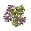
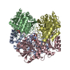
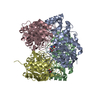
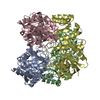
| Title | Wolinella succinogenes L-asparaginase S121 + L-aspartic acid, open conformation | ||||||
 Components Components | L-asparaginase | ||||||
 Keywords Keywords | HYDROLASE / S121 / L-aspartic acid / open conformation | ||||||
| Function / homology |  Function and homology information Function and homology information | ||||||
| Biological species |  Wolinella succinogenes (bacteria) Wolinella succinogenes (bacteria) | ||||||
| Method |  X-RAY DIFFRACTION / SYNCHROTRON / MOLECULAR REPLACEMENT / Resolution: 1.6 Å X-RAY DIFFRACTION / SYNCHROTRON / MOLECULAR REPLACEMENT / Resolution: 1.6 Å | ||||||
 Authors Authors | Nguyen, H.A. / Lave, A. | ||||||
 Citation Citation | Journal: Sci Rep / Year: 2017 Title: The differential ability of asparagine and glutamine in promoting the closed/active enzyme conformation rationalizes the Wolinella succinogenes L-asparaginase substrate specificity. Authors: Nguyen, H.A. / Durden, D.L. / Lavie, A. #1: Journal: Biochemistry / Year: 2016Title: Structural Insight into Substrate Selectivity of Erwinia chrysanthemi l-Asparaginase. Authors: Nguyen, H.A. / Su, Y. / Lavie, A. | ||||||
| History |
|
- Structure visualization
| Structure viewer | Molecule: MolmilJmol/JSmol |
|---|
- Downloads & links
-Download
| PDBx/mmCIF format | 5k4g.cif.gz | 282.7 KB | Display | PDBx/mmCIF format |
|---|---|---|---|---|
| PDB format | pdb5k4g.ent.gz | 225.4 KB | Display | PDB format |
| PDBx/mmJSON format | 5k4g.json.gz | Tree view | PDBx/mmJSON format | |
| Others |  Other downloads Other downloads |
-Validation report
| Arichive directory | https://data.pdbj.org/pub/pdb/validation_reports/k4/5k4gftp://data.pdbj.org/pub/pdb/validation_reports/k4/5k4g | HTTPS FTP |
|---|
-Related structure data
| Related structure data |  5k3oC  5k45C  5k4hC  1wsaS C: citing same article ( S: Starting model for refinement |
|---|---|
| Similar structure data |

-Links
 PDBj
PDBj
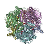
- Assembly
| Deposited unit | 
| ||||||||||||||||||||||||||||||||||||||||||||||||||||||||||||||||||||||||||||||||||||||||||||||||||
|---|---|---|---|---|---|---|---|---|---|---|---|---|---|---|---|---|---|---|---|---|---|---|---|---|---|---|---|---|---|---|---|---|---|---|---|---|---|---|---|---|---|---|---|---|---|---|---|---|---|---|---|---|---|---|---|---|---|---|---|---|---|---|---|---|---|---|---|---|---|---|---|---|---|---|---|---|---|---|---|---|---|---|---|---|---|---|---|---|---|---|---|---|---|---|---|---|---|---|---|
| 1 | 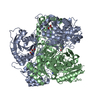
| ||||||||||||||||||||||||||||||||||||||||||||||||||||||||||||||||||||||||||||||||||||||||||||||||||
| 2 | 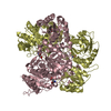
| ||||||||||||||||||||||||||||||||||||||||||||||||||||||||||||||||||||||||||||||||||||||||||||||||||
| Unit cell |
| ||||||||||||||||||||||||||||||||||||||||||||||||||||||||||||||||||||||||||||||||||||||||||||||||||
| Noncrystallographic symmetry (NCS) | NCS domain:
NCS domain segments: Component-ID: _ / Beg auth comp-ID: LYS / Beg label comp-ID: LYS / End auth comp-ID: TYR / End label comp-ID: TYR / Refine code: _ / Auth seq-ID: 3 - 330 / Label seq-ID: 3 - 330
NCS ensembles :
|
-Components
| #1: Protein | Mass: 34893.703 Da / Num. of mol.: 4 Source method: isolated from a genetically manipulated source Source: (gene. exp.) Wolinella succinogenes (strain ATCC 29543 / DSM 1740 / LMG 7466 / NCTC 11488 / FDC 602W) (bacteria)Strain: ATCC 29543 / DSM 1740 / LMG 7466 / NCTC 11488 / FDC 602W Gene: ansA, ansB, WS0660 / Production host: #2: Chemical | ChemComp-ASP /   Type: L-peptide linking / Mass: 133.103 Da / Num. of mol.: 4 / Source method: obtained synthetically / Formula: C4H7NO4 Type: L-peptide linking / Mass: 133.103 Da / Num. of mol.: 4 / Source method: obtained synthetically / Formula: C4H7NO4#3: Water | ChemComp-HOH / |  Mass: 18.015 Da / Num. of mol.: 1367 / Source method: isolated from a natural source / Formula: H2O Mass: 18.015 Da / Num. of mol.: 1367 / Source method: isolated from a natural source / Formula: H2O |
|---|
-Experimental details
-Experiment
| Experiment | Method: X-RAY DIFFRACTION / Number of used crystals: 1 |
|---|
- Sample preparation
| Crystal | Density Matthews: 2.36 Å3/Da / Density % sol: 47.87 % |
|---|---|
| Crystal grow | Temperature: 283 K / Method: vapor diffusion, hanging drop / pH: 7.5 / Details: PEG2000, HEPES 7.5 |
-Data collection
| Diffraction | Mean temperature: 100 K | ||||||||||||||||||||||||||||||||||||||||||||||||||||||||||||
|---|---|---|---|---|---|---|---|---|---|---|---|---|---|---|---|---|---|---|---|---|---|---|---|---|---|---|---|---|---|---|---|---|---|---|---|---|---|---|---|---|---|---|---|---|---|---|---|---|---|---|---|---|---|---|---|---|---|---|---|---|---|
| Diffraction source | Source: SYNCHROTRON / Site: APS  / Beamline: 21-ID-G / Wavelength: 0.97857 Å / Beamline: 21-ID-G / Wavelength: 0.97857 Å | ||||||||||||||||||||||||||||||||||||||||||||||||||||||||||||
| Detector | Type: MARMOSAIC 300 mm CCD / Detector: CCD / Date: Oct 17, 2015 | ||||||||||||||||||||||||||||||||||||||||||||||||||||||||||||
| Radiation | Protocol: MAD / Monochromatic (M) / Laue (L): M / Scattering type: x-ray | ||||||||||||||||||||||||||||||||||||||||||||||||||||||||||||
| Radiation wavelength | Wavelength: 0.97857 Å / Relative weight: 1 | ||||||||||||||||||||||||||||||||||||||||||||||||||||||||||||
| Reflection | Resolution: 1.6→30 Å / Num. obs: 166478 / % possible obs: 97.5 % / Observed criterion σ(I): -3 / Redundancy: 3.14 % / Biso Wilson estimate: 25.71 Å2 / CC1/2: 0.999 / Rmerge(I) obs: 0.04 / Net I/σ(I): 17.99 | ||||||||||||||||||||||||||||||||||||||||||||||||||||||||||||
| Reflection shell |
|
- Processing
| Software |
| |||||||||||||||||||||||||||||||||||||||||||||||||||||||||||||||||||||||||||
|---|---|---|---|---|---|---|---|---|---|---|---|---|---|---|---|---|---|---|---|---|---|---|---|---|---|---|---|---|---|---|---|---|---|---|---|---|---|---|---|---|---|---|---|---|---|---|---|---|---|---|---|---|---|---|---|---|---|---|---|---|---|---|---|---|---|---|---|---|---|---|---|---|---|---|---|---|
| Refinement | Method to determine structure: MOLECULAR REPLACEMENT Starting model: 1WSA Resolution: 1.6→30 Å / Cor.coef. Fo:Fc: 0.972 / Cor.coef. Fo:Fc free: 0.957 / SU B: 2.109 / SU ML: 0.07 / SU R Cruickshank DPI: 0.0867 / Cross valid method: THROUGHOUT / σ(F): 0 / ESU R: 0.087 / ESU R Free: 0.089 Details: HYDROGENS HAVE BEEN ADDED IN THE RIDING POSITIONS U VALUES : REFINED INDIVIDUALLY
| |||||||||||||||||||||||||||||||||||||||||||||||||||||||||||||||||||||||||||
| Solvent computation | Ion probe radii: 0.8 Å / Shrinkage radii: 0.8 Å / VDW probe radii: 1.2 Å | |||||||||||||||||||||||||||||||||||||||||||||||||||||||||||||||||||||||||||
| Displacement parameters | Biso max: 97.93 Å2 / Biso mean: 23.727 Å2 / Biso min: 8.63 Å2
| |||||||||||||||||||||||||||||||||||||||||||||||||||||||||||||||||||||||||||
| Refinement step | Cycle: final / Resolution: 1.6→30 Å
| |||||||||||||||||||||||||||||||||||||||||||||||||||||||||||||||||||||||||||
| Refine LS restraints |
| |||||||||||||||||||||||||||||||||||||||||||||||||||||||||||||||||||||||||||
| Refine LS restraints NCS | Refine-ID: X-RAY DIFFRACTION / Type: interatomic distance / Weight position: 0.05
| |||||||||||||||||||||||||||||||||||||||||||||||||||||||||||||||||||||||||||
| LS refinement shell | Resolution: 1.599→1.641 Å / Total num. of bins used: 20
|
