 Movie
Movie Controller

[English] 日本語
 Yorodumi
- PDB-3uob: Crystal structure of Human Thymine DNA Glycosylase Bound to Subst... -
+ Open data

- Basic information
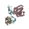
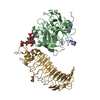
| Entry | Database: PDB / ID: 3uob | ||||||
|---|---|---|---|---|---|---|---|
| Title | Crystal structure of Human Thymine DNA Glycosylase Bound to Substrate Analog 2'-deoxy-2'-beta-fluoro-cytidine | ||||||
 Components Components |
| ||||||
 Keywords Keywords | HYDROLASE/DNA / dsDNA / HYDROLASE-DNA complex | ||||||
| Function / homology |  Function and homology information Function and homology informationG/U mismatch-specific uracil-DNA glycosylase activity / thymine-DNA glycosylase / G/T mismatch-specific thymine-DNA glycosylase activity / chromosomal 5-methylcytosine DNA demethylation, oxidation pathway / TET1,2,3 and TDG demethylate DNA / pyrimidine-specific mismatch base pair DNA N-glycosylase activity / base-excision repair, AP site formation / depyrimidination / DNA N-glycosylase activity / sodium ion binding ...G/U mismatch-specific uracil-DNA glycosylase activity / thymine-DNA glycosylase / G/T mismatch-specific thymine-DNA glycosylase activity / chromosomal 5-methylcytosine DNA demethylation, oxidation pathway / TET1,2,3 and TDG demethylate DNA / pyrimidine-specific mismatch base pair DNA N-glycosylase activity / base-excision repair, AP site formation / depyrimidination / DNA N-glycosylase activity / sodium ion binding / mismatched DNA binding / Displacement of DNA glycosylase by APEX1 / SUMO binding / uracil DNA N-glycosylase activity / chloride ion binding / regulation of embryonic development / SUMOylation of DNA damage response and repair proteins / epigenetic regulation of gene expression / Recognition and association of DNA glycosylase with site containing an affected pyrimidine / Cleavage of the damaged pyrimidine / protein kinase C binding / transcription coregulator activity / base-excision repair / PML body / double-stranded DNA binding / nucleic acid binding / damaged DNA binding / magnesium ion binding / DNA-templated transcription / DNA binding / nucleoplasm / ATP binding / nucleus Similarity search - Function | ||||||
| Biological species |  Homo sapiens (human) Homo sapiens (human) | ||||||
| Method |  X-RAY DIFFRACTION / SYNCHROTRON / MOLECULAR REPLACEMENT / Resolution: 3.011 Å X-RAY DIFFRACTION / SYNCHROTRON / MOLECULAR REPLACEMENT / Resolution: 3.011 Å | ||||||
 Authors Authors | Zhang, L. / He, C. | ||||||
 Citation Citation | Journal: Nat.Chem.Biol. / Year: 2012 Title: Thymine DNA glycosylase specifically recognizes 5-carboxylcytosine-modified DNA. Authors: Zhang, L. / Lu, X. / Lu, J. / Liang, H. / Dai, Q. / Xu, G.L. / Luo, C. / Jiang, H. / He, C. | ||||||
| History |
|
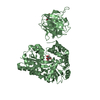
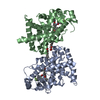
- Structure visualization
| Structure viewer | Molecule: MolmilJmol/JSmol |
|---|
- Downloads & links
-Download
| PDBx/mmCIF format | 3uob.cif.gz | 111.4 KB | Display | PDBx/mmCIF format |
|---|---|---|---|---|
| PDB format | pdb3uob.ent.gz | 82.6 KB | Display | PDB format |
| PDBx/mmJSON format | 3uob.json.gz | Tree view | PDBx/mmJSON format | |
| Others |  Other downloads Other downloads |
-Validation report
| Arichive directory | https://data.pdbj.org/pub/pdb/validation_reports/uo/3uobftp://data.pdbj.org/pub/pdb/validation_reports/uo/3uob | HTTPS FTP |
|---|
-Related structure data
| Related structure data |  3uo7C  2rbaS C: citing same article ( S: Starting model for refinement |
|---|---|
| Similar structure data |


-Links
 PDBj
PDBj


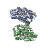
- Assembly
| Deposited unit | 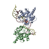
| ||||||||
|---|---|---|---|---|---|---|---|---|---|
| 1 |
| ||||||||
| Unit cell |
|
-Components
| #1: DNA chain | Mass: 7121.597 Da / Num. of mol.: 1 / Source method: obtained synthetically |
|---|---|
| #2: DNA chain | Mass: 7063.524 Da / Num. of mol.: 1 / Source method: obtained synthetically |
| #3: Protein | Mass: 22771.301 Da / Num. of mol.: 2 / Fragment: UNP residues 111-308 Source method: isolated from a genetically manipulated source Source: (gene. exp.) Homo sapiens (human) / Gene: TDG / Production host:  |
-Experimental details
-Experiment
| Experiment | Method: X-RAY DIFFRACTION / Number of used crystals: 1 |
|---|
- Sample preparation
| Crystal | Density Matthews: 3.53 Å3/Da / Density % sol: 65.16 % |
|---|---|
| Crystal grow | Temperature: 293 K / Method: vapor diffusion, hanging drop / pH: 7 Details: 25% PEG3350, 0.2 M tripotassium citrate monohydrate, pH 7.0, VAPOR DIFFUSION, HANGING DROP, temperature 293K |
-Data collection
| Diffraction | Mean temperature: 180 K | |||||||||||||||||||||||||||||||||||
|---|---|---|---|---|---|---|---|---|---|---|---|---|---|---|---|---|---|---|---|---|---|---|---|---|---|---|---|---|---|---|---|---|---|---|---|---|
| Diffraction source | Source: SYNCHROTRON / Site: APS  / Beamline: 24-ID-E / Wavelength: 0.97918 Å / Beamline: 24-ID-E / Wavelength: 0.97918 Å | |||||||||||||||||||||||||||||||||||
| Detector | Type: ADSC QUANTUM 315 / Detector: CCD / Date: Oct 14, 2011 | |||||||||||||||||||||||||||||||||||
| Radiation | Monochromator: Cryogenically-cooled single crystal Si(220) side bounce Protocol: SINGLE WAVELENGTH / Monochromatic (M) / Laue (L): M / Scattering type: x-ray | |||||||||||||||||||||||||||||||||||
| Radiation wavelength | Wavelength: 0.97918 Å / Relative weight: 1 | |||||||||||||||||||||||||||||||||||
| Reflection | Resolution: 3→50 Å / Num. all: 11703 / Num. obs: 11703 / % possible obs: 59.3 % / Observed criterion σ(F): 2 / Observed criterion σ(I): 1 / Redundancy: 4.7 % / Rmerge(I) obs: 0.096 / Rsym value: 0.096 / Net I/σ(I): 13 | |||||||||||||||||||||||||||||||||||
| Reflection shell |
|
- Processing
| Software |
| |||||||||||||||||||||||||||||||||||||||||||||||||||||||||||||||
|---|---|---|---|---|---|---|---|---|---|---|---|---|---|---|---|---|---|---|---|---|---|---|---|---|---|---|---|---|---|---|---|---|---|---|---|---|---|---|---|---|---|---|---|---|---|---|---|---|---|---|---|---|---|---|---|---|---|---|---|---|---|---|---|---|
| Refinement | Method to determine structure: MOLECULAR REPLACEMENT Starting model: PDB ENTRY 2RBA Resolution: 3.011→32.733 Å / SU ML: 0.99 / σ(F): 1.35 / Phase error: 32.74 / Stereochemistry target values: ML
| |||||||||||||||||||||||||||||||||||||||||||||||||||||||||||||||
| Solvent computation | Shrinkage radii: 0.86 Å / VDW probe radii: 1.1 Å / Solvent model: FLAT BULK SOLVENT MODEL / Bsol: 45.407 Å2 / ksol: 0.272 e/Å3 | |||||||||||||||||||||||||||||||||||||||||||||||||||||||||||||||
| Displacement parameters |
| |||||||||||||||||||||||||||||||||||||||||||||||||||||||||||||||
| Refinement step | Cycle: LAST / Resolution: 3.011→32.733 Å
| |||||||||||||||||||||||||||||||||||||||||||||||||||||||||||||||
| Refine LS restraints |
| |||||||||||||||||||||||||||||||||||||||||||||||||||||||||||||||
| LS refinement shell |
|
