 Movie
Movie Controller


+ Open data

- Basic information
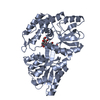
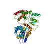
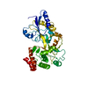
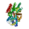
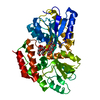
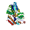
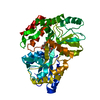
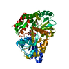
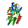
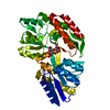
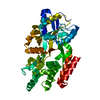
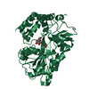
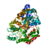
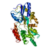
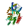
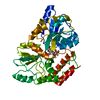
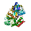
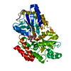
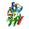
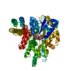
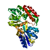
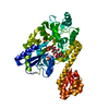
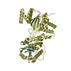
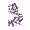
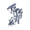
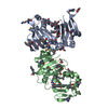
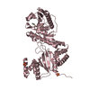
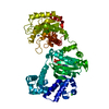
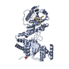
| Entry | Database: PDB / ID: 4b3n | |||||||||
|---|---|---|---|---|---|---|---|---|---|---|
| Title | Crystal structure of rhesus TRIM5alpha PRY/SPRY domain | |||||||||
 Components Components | MALTOSE-BINDING PERIPLASMIC PROTEIN, TRIPARTITE MOTIF-CONTAINING PROTEIN 5 | |||||||||
 Keywords Keywords | SUGAR BINDING PROTEIN/LIGASE / SUGAR BINDING PROTEIN-LIGASE COMPLEX / SUGAR BINDING PROTEIN-LIGASE CHIMERA | |||||||||
| Function / homology |  Function and homology information Function and homology informationregulation of viral entry into host cell / regulation of lipopolysaccharide-mediated signaling pathway / host-mediated suppression of symbiont invasion / suppression of viral release by host / negative regulation of viral transcription / negative regulation of viral genome replication / pattern recognition receptor activity / detection of maltose stimulus / maltose transport complex / carbohydrate transport ...regulation of viral entry into host cell / regulation of lipopolysaccharide-mediated signaling pathway / host-mediated suppression of symbiont invasion / suppression of viral release by host / negative regulation of viral transcription / negative regulation of viral genome replication / pattern recognition receptor activity / detection of maltose stimulus / maltose transport complex / carbohydrate transport / carbohydrate transmembrane transporter activity / maltose binding / maltose transport / maltodextrin transmembrane transport / protein K63-linked ubiquitination / ATP-binding cassette (ABC) transporter complex, substrate-binding subunit-containing / activation of innate immune response / ATP-binding cassette (ABC) transporter complex / : / cell chemotaxis / P-body / RING-type E3 ubiquitin transferase / autophagy / ubiquitin-protein transferase activity / ubiquitin protein ligase activity / regulation of protein localization / outer membrane-bounded periplasmic space / regulation of gene expression / defense response to virus / periplasmic space / positive regulation of canonical NF-kappaB signal transduction / positive regulation of MAPK cascade / innate immune response / DNA damage response / protein kinase binding / protein homodimerization activity / zinc ion binding / membrane / identical protein binding / nucleus / cytoplasm Similarity search - Function | |||||||||
| Biological species |   | |||||||||
| Method |  X-RAY DIFFRACTION / SYNCHROTRON / MOLECULAR REPLACEMENT / Resolution: 3.3 Å X-RAY DIFFRACTION / SYNCHROTRON / MOLECULAR REPLACEMENT / Resolution: 3.3 Å | |||||||||
 Authors Authors | Yang, H. / Ji, X. / Zhao, Q. / Xiong, Y. | |||||||||
 Citation Citation | Journal: Proc.Natl.Acad.Sci.USA / Year: 2012 Title: Structural Insight Into HIV-1 Capsid Recognition by Rhesus Trim5Alpha Authors: Yang, H. / Ji, X. / Zhao, G. / Ning, J. / Zhao, Q. / Aiken, C. / Gronenborn, A.M. / Zhang, P. / Xiong, Y. | |||||||||
| History |
|
- Structure visualization
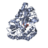
| Structure viewer | Molecule: MolmilJmol/JSmol |
|---|
- Downloads & links
-Download
| PDBx/mmCIF format | 4b3n.cif.gz | 459.2 KB | Display | PDBx/mmCIF format |
|---|---|---|---|---|
| PDB format | pdb4b3n.ent.gz | 380.5 KB | Display | PDB format |
| PDBx/mmJSON format | 4b3n.json.gz | Tree view | PDBx/mmJSON format | |
| Others |  Other downloads Other downloads |
-Validation report
| Arichive directory | https://data.pdbj.org/pub/pdb/validation_reports/b3/4b3nftp://data.pdbj.org/pub/pdb/validation_reports/b3/4b3n | HTTPS FTP |
|---|
-Related structure data
| Related structure data |  1anfS  2vol 4auf 4aug 4auh S: Starting model for refinement |
|---|---|
| Similar structure data |


-Links
 PDBj
PDBj


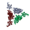
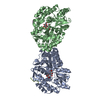
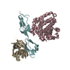
- Assembly
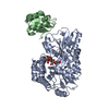
| Deposited unit | 
| ||||||||
|---|---|---|---|---|---|---|---|---|---|
| 1 | 
| ||||||||
| 2 | 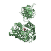
| ||||||||
| Unit cell |
|
-Components
| #1: Protein | Mass: 67269.273 Da / Num. of mol.: 2 Fragment: MBP RESIDUES 27-395, TRIM5ALPHA PRY/SPRY DOMAIN RESIDUES 275-493 Source method: isolated from a genetically manipulated source Source: (gene. exp.) Plasmid: PMAT9S / Production host: #2: Polysaccharide |   Source method: isolated from a genetically manipulated source Details: oligosaccharide / References: alpha-maltose #3: Chemical |   Mass: 195.237 Da / Num. of mol.: 2 / Source method: obtained synthetically / Formula: C6H13NO4S / Comment: pH buffer*YM Mass: 195.237 Da / Num. of mol.: 2 / Source method: obtained synthetically / Formula: C6H13NO4S / Comment: pH buffer*YM#4: Water | ChemComp-HOH / |  Mass: 18.015 Da / Num. of mol.: 44 / Source method: isolated from a natural source / Formula: H2O Mass: 18.015 Da / Num. of mol.: 44 / Source method: isolated from a natural source / Formula: H2O |
|---|
-Experimental details
-Experiment
| Experiment | Method: X-RAY DIFFRACTION / Number of used crystals: 4 |
|---|
- Sample preparation
| Crystal | Density Matthews: 2.9 Å3/Da / Density % sol: 59 % / Description: NONE |
|---|---|
| Crystal grow | pH: 6.2 Details: 6% (W/V) GLUCOSE, 6% (W/V) TREHALOSE, 100 MM MES PH 6.2 AND 25% PEG 3350 |
-Data collection
| Diffraction | Mean temperature: 100 K |
|---|---|
| Diffraction source | Source: SYNCHROTRON / Site: NSLS  / Beamline: X29A / Wavelength: 1.075 / Beamline: X29A / Wavelength: 1.075 |
| Detector | Type: ADSC CCD / Detector: CCD / Date: Apr 30, 2012 Details: HORIZONTAL FOCUSING SAGITTAL BEND SECOND MONO CRYSTAL AND VERTICALLY FOCUSING MIRROR |
| Radiation | Monochromator: CRYOGENICALLY COOLED DOUBLE CRYSTAL MONOCHROMETER Protocol: SINGLE WAVELENGTH / Monochromatic (M) / Laue (L): M / Scattering type: x-ray |
| Radiation wavelength | Wavelength: 1.075 Å / Relative weight: 1 |
| Reflection | Resolution: 3.3→50 Å / Num. obs: 22400 / % possible obs: 96.5 % / Observed criterion σ(I): 1 / Redundancy: 3 % / Rmerge(I) obs: 0.17 / Net I/σ(I): 6.69 |
| Reflection shell | Resolution: 3.3→3.42 Å / Redundancy: 3 % / Rmerge(I) obs: 0.89 / Mean I/σ(I) obs: 1.39 / % possible all: 97.1 |
- Processing
| Software |
| ||||||||||||||||||||||||||||||||||||||||||||||||||||||||||||||||||||||||||||||||||||||||||||||||||||||||||||||||||||||||||||||||||||||||||||||||||||||||||||||||||||||||||||||||||||||
|---|---|---|---|---|---|---|---|---|---|---|---|---|---|---|---|---|---|---|---|---|---|---|---|---|---|---|---|---|---|---|---|---|---|---|---|---|---|---|---|---|---|---|---|---|---|---|---|---|---|---|---|---|---|---|---|---|---|---|---|---|---|---|---|---|---|---|---|---|---|---|---|---|---|---|---|---|---|---|---|---|---|---|---|---|---|---|---|---|---|---|---|---|---|---|---|---|---|---|---|---|---|---|---|---|---|---|---|---|---|---|---|---|---|---|---|---|---|---|---|---|---|---|---|---|---|---|---|---|---|---|---|---|---|---|---|---|---|---|---|---|---|---|---|---|---|---|---|---|---|---|---|---|---|---|---|---|---|---|---|---|---|---|---|---|---|---|---|---|---|---|---|---|---|---|---|---|---|---|---|---|---|---|---|
| Refinement | Method to determine structure: MOLECULAR REPLACEMENT Starting model: PDB ENTRIES 1ANF, 2VOL Resolution: 3.3→49.42 Å / Cor.coef. Fo:Fc: 0.923 / Cor.coef. Fo:Fc free: 0.875 / SU B: 59.229 / SU ML: 0.429 / Cross valid method: THROUGHOUT / ESU R Free: 0.546 / Stereochemistry target values: MAXIMUM LIKELIHOOD Details: HYDROGENS HAVE BEEN ADDED IN THE RIDING POSITIONS. U VALUES WITH TLS ADDED
| ||||||||||||||||||||||||||||||||||||||||||||||||||||||||||||||||||||||||||||||||||||||||||||||||||||||||||||||||||||||||||||||||||||||||||||||||||||||||||||||||||||||||||||||||||||||
| Solvent computation | Ion probe radii: 0.8 Å / Shrinkage radii: 0.86 Å / VDW probe radii: 1.1 Å / Solvent model: MASK | ||||||||||||||||||||||||||||||||||||||||||||||||||||||||||||||||||||||||||||||||||||||||||||||||||||||||||||||||||||||||||||||||||||||||||||||||||||||||||||||||||||||||||||||||||||||
| Displacement parameters | Biso mean: 85.292 Å2
| ||||||||||||||||||||||||||||||||||||||||||||||||||||||||||||||||||||||||||||||||||||||||||||||||||||||||||||||||||||||||||||||||||||||||||||||||||||||||||||||||||||||||||||||||||||||
| Refinement step | Cycle: LAST / Resolution: 3.3→49.42 Å
| ||||||||||||||||||||||||||||||||||||||||||||||||||||||||||||||||||||||||||||||||||||||||||||||||||||||||||||||||||||||||||||||||||||||||||||||||||||||||||||||||||||||||||||||||||||||
| Refine LS restraints |
|