 Movie
Movie Controller

[English] 日本語
 Yorodumi
- PDB-4wvg: Crystal structure of the Type-I signal peptidase from Staphylococ... -
+ Open data

- Basic information
| Entry | Database: PDB / ID: 4wvg | |||||||||
|---|---|---|---|---|---|---|---|---|---|---|
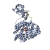
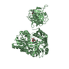
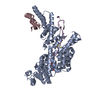
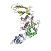
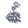
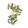
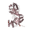
| Title | Crystal structure of the Type-I signal peptidase from Staphylococcus aureus (SpsB). | |||||||||
 Components Components | Maltose-binding periplasmic protein,Signal peptidase IB | |||||||||
 Keywords Keywords | HYDROLASE / SpsB Type-I signal peptidase / Cell secretion / MBP fusion protein | |||||||||
| Function / homology |  Function and homology information Function and homology informationsignal peptidase I / signal peptidase activity / : / carbohydrate transmembrane transporter activity / maltose binding / maltose transport / maltodextrin transmembrane transport / ATP-binding cassette (ABC) transporter complex, substrate-binding subunit-containing / outer membrane-bounded periplasmic space / serine-type endopeptidase activity / plasma membrane Similarity search - Function | |||||||||
| Biological species |  Staphylococcus aureus subsp. aureus str. Newman (bacteria) Staphylococcus aureus subsp. aureus str. Newman (bacteria) | |||||||||
| Method |  X-RAY DIFFRACTION / SYNCHROTRON / MOLECULAR REPLACEMENT / molecular replacement / Resolution: 2.05 Å X-RAY DIFFRACTION / SYNCHROTRON / MOLECULAR REPLACEMENT / molecular replacement / Resolution: 2.05 Å | |||||||||
 Authors Authors | Young, P.G. / Ting, Y.T. / Baker, E.N. | |||||||||
| Funding support |  New Zealand, 1items New Zealand, 1items
| |||||||||
 Citation Citation | Journal: IUCrJ / Year: 2016 Title: Peptide binding to a bacterial signal peptidase visualized by peptide tethering and carrier-driven crystallization. Authors: Ting, Y.T. / Harris, P.W. / Batot, G. / Brimble, M.A. / Baker, E.N. / Young, P.G. | |||||||||
| History |
|
- Structure visualization
| Structure viewer | Molecule: MolmilJmol/JSmol |
|---|
- Downloads & links
-Download
| PDBx/mmCIF format | 4wvg.cif.gz | 124.2 KB | Display | PDBx/mmCIF format |
|---|---|---|---|---|
| PDB format | pdb4wvg.ent.gz | 92.5 KB | Display | PDB format |
| PDBx/mmJSON format | 4wvg.json.gz | Tree view | PDBx/mmJSON format | |
| Others |  Other downloads Other downloads |
-Validation report
| Arichive directory | https://data.pdbj.org/pub/pdb/validation_reports/wv/4wvgftp://data.pdbj.org/pub/pdb/validation_reports/wv/4wvg | HTTPS FTP |
|---|
-Related structure data
| Related structure data |  4wvhC  4wviC 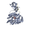 4wvjC  1anfS C: citing same article ( S: Starting model for refinement |
|---|---|
| Similar structure data |


-Links
 PDBj
PDBj


- Assembly
| Deposited unit | 
| ||||||||
|---|---|---|---|---|---|---|---|---|---|
| 1 |
| ||||||||
| Unit cell |
|
-Components
| #1: Protein | Mass: 60301.949 Da / Num. of mol.: 1 / Fragment: UNP RESIDUES 33-393, UNP RESIDUES 26-191 / Mutation: S36A, R393N Source method: isolated from a genetically manipulated source Source: (gene. exp.) Staphylococcus aureus subsp. aureus str. Newman (bacteria)Strain: K12, Newman / Gene: malE, Z5632, ECs5017, spsB, SACOL0969 / Plasmid: pMBP-pProExHta / Details (production host): MBP fusion / Production host: References: UniProt: P0AEY0, UniProt: Q5HHB9, signal peptidase I |
|---|---|
| #2: Polysaccharide | alpha-D-glucopyranose-(1-4)-alpha-D-glucopyranose / alpha-maltose  Source method: isolated from a genetically manipulated source Details: oligosaccharide / References: alpha-maltose |
| #3: Water | ChemComp-HOH /  Mass: 18.015 Da / Num. of mol.: 356 / Source method: isolated from a natural source / Formula: H2O Mass: 18.015 Da / Num. of mol.: 356 / Source method: isolated from a natural source / Formula: H2O |
| Sequence details | THIS MUTATION IS A RESULT OF PCR REACTION. THE MUTATION R393N HAS BEEN INTRODUCED TO STABILIZE THE ...THIS MUTATION IS A RESULT OF PCR REACTION. THE MUTATION R393N HAS BEEN INTRODUCED |
-Experimental details
-Experiment
| Experiment | Method: X-RAY DIFFRACTION / Number of used crystals: 1 |
|---|
- Sample preparation
| Crystal | Density Matthews: 2.43 Å3/Da / Density % sol: 49.31 % / Description: Needles |
|---|---|
| Crystal grow | Temperature: 296 K / Method: vapor diffusion, hanging drop Details: 12 % PEG 8000, 20 % ethylene glycol, 0.2 M amino acids mix (0.2 M sodium-L-glutamate, 0.2 M DL-alanine, 0.2 M glycine, 0.2 M DL-lysine, 0.2 M DL-serine), 100 mM Tris.Cl pH 8.5 PH range: 8.0 - 8.5 |
-Data collection
| Diffraction | Mean temperature: 100 K | |||||||||||||||||||||||||||
|---|---|---|---|---|---|---|---|---|---|---|---|---|---|---|---|---|---|---|---|---|---|---|---|---|---|---|---|---|
| Diffraction source | Source: SYNCHROTRON / Site: Australian Synchrotron  / Beamline: MX1 / Wavelength: 0.9537 Å / Beamline: MX1 / Wavelength: 0.9537 Å | |||||||||||||||||||||||||||
| Detector | Type: ADSC QUANTUM 210r / Detector: CCD / Date: Mar 25, 2013 | |||||||||||||||||||||||||||
| Radiation | Protocol: SINGLE WAVELENGTH / Monochromatic (M) / Laue (L): M / Scattering type: x-ray | |||||||||||||||||||||||||||
| Radiation wavelength | Wavelength: 0.9537 Å / Relative weight: 1 | |||||||||||||||||||||||||||
| Reflection | Resolution: 2.05→19.44 Å / Num. obs: 36318 / % possible obs: 99.8 % / Redundancy: 7.6 % / CC1/2: 0.994 / Rmerge(I) obs: 0.211 / Rpim(I) all: 0.082 / Net I/σ(I): 10.1 / Num. measured all: 275184 | |||||||||||||||||||||||||||
| Reflection shell | Diffraction-ID: 1 / Rejects: _
|
-Phasing
| Phasing | Method: molecular replacement |
|---|
- Processing
| Software |
| |||||||||||||||||||||||||||||||||||||||||||||||||||||||||||||||||||||||||||
|---|---|---|---|---|---|---|---|---|---|---|---|---|---|---|---|---|---|---|---|---|---|---|---|---|---|---|---|---|---|---|---|---|---|---|---|---|---|---|---|---|---|---|---|---|---|---|---|---|---|---|---|---|---|---|---|---|---|---|---|---|---|---|---|---|---|---|---|---|---|---|---|---|---|---|---|---|
| Refinement | Method to determine structure: MOLECULAR REPLACEMENT Starting model: 1ANF Resolution: 2.05→19.44 Å / Cor.coef. Fo:Fc: 0.954 / Cor.coef. Fo:Fc free: 0.923 / WRfactor Rfree: 0.1901 / WRfactor Rwork: 0.1481 / FOM work R set: 0.7898 / SU B: 5.971 / SU ML: 0.151 / SU R Cruickshank DPI: 0.2001 / SU Rfree: 0.1796 / Cross valid method: THROUGHOUT / σ(F): 0 / ESU R: 0.2 / ESU R Free: 0.18 / Stereochemistry target values: MAXIMUM LIKELIHOOD Details: HYDROGENS HAVE BEEN ADDED IN THE RIDING POSITIONS U VALUES : REFINED INDIVIDUALLY
| |||||||||||||||||||||||||||||||||||||||||||||||||||||||||||||||||||||||||||
| Solvent computation | Ion probe radii: 0.8 Å / Shrinkage radii: 0.8 Å / VDW probe radii: 1.2 Å / Solvent model: MASK | |||||||||||||||||||||||||||||||||||||||||||||||||||||||||||||||||||||||||||
| Displacement parameters | Biso max: 64.54 Å2 / Biso mean: 29.325 Å2 / Biso min: 14.23 Å2
| |||||||||||||||||||||||||||||||||||||||||||||||||||||||||||||||||||||||||||
| Refinement step | Cycle: final / Resolution: 2.05→19.44 Å
| |||||||||||||||||||||||||||||||||||||||||||||||||||||||||||||||||||||||||||
| Refine LS restraints |
| |||||||||||||||||||||||||||||||||||||||||||||||||||||||||||||||||||||||||||
| LS refinement shell | Resolution: 2.05→2.103 Å / Total num. of bins used: 20
|
