| Entry | Database: PDB / ID: 6u8c
|
|---|
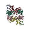
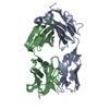
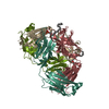
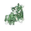
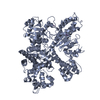
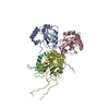
| Title | Crystal structure of an engineered ultra-high affinity Fab-Protein G complex |
|---|
 Components Components | - Antibody heavy chain Fab
- Antibody light chain Fab
- Protein G
|
|---|
 Keywords Keywords | IMMUNE SYSTEM / engineered Fab / protein G / high affinity |
|---|
| Function / homology |  Function and homology information Function and homology information
Ubiquitin-like (UB roll) - #10 / IgG-binding B / B domain / M protein-type anchor domain / GA-like domain / GA-like domain / Immunoglobulin/albumin-binding domain superfamily / YSIRK Gram-positive signal peptide / LPXTG cell wall anchor motif / Gram-positive cocci surface proteins LPxTG motif profile. ...Ubiquitin-like (UB roll) - #10 / IgG-binding B / B domain / M protein-type anchor domain / GA-like domain / GA-like domain / Immunoglobulin/albumin-binding domain superfamily / YSIRK Gram-positive signal peptide / LPXTG cell wall anchor motif / Gram-positive cocci surface proteins LPxTG motif profile. / LPXTG cell wall anchor domain / Ubiquitin-like (UB roll) / Roll / Immunoglobulins / Immunoglobulin-like / Sandwich / Mainly Beta / Alpha BetaSimilarity search - Domain/homology |
|---|
| Biological species |  Streptococcus sp. 'group G' (bacteria) Streptococcus sp. 'group G' (bacteria)
 Homo sapiens (human) Homo sapiens (human) |
|---|
| Method |  X-RAY DIFFRACTION / SYNCHROTRON / MOLECULAR REPLACEMENT / Resolution: 2.61 Å X-RAY DIFFRACTION / SYNCHROTRON / MOLECULAR REPLACEMENT / Resolution: 2.61 Å |
|---|
 Authors Authors | Slezak, T. / Filippova, E.V. / Davydova, E.K. / Kossiakoff, A.A. |
|---|
| Funding support |  United States, 1items United States, 1items | Organization | Grant number | Country |
|---|
| National Institutes of Health/National Institute of Dental and Craniofacial Research (NIH/NIDCR) | GM117372 | United States |
|
|---|
 Citation Citation | Journal: Protein Sci. / Year: 2020
Title: An engineered ultra-high affinity Fab-Protein G pair enables a modular antibody platform with multifunctional capability.
Authors: Slezak, T. / Bailey, L.J. / Jaskolowski, M. / Nahotko, D.A. / Filippova, E.V. / Davydova, E.K. / Kossiakoff, A.A. |
|---|
| History | | Deposition | Sep 4, 2019 | Deposition site: RCSB / Processing site: RCSB |
|---|
| Revision 1.0 | Oct 23, 2019 | Provider: repository / Type: Initial release |
|---|
| Revision 1.1 | Oct 30, 2019 | Group: Data collection / Database references / Category: citation / Item: _citation.pdbx_database_id_PubMed / _citation.title |
|---|
| Revision 1.2 | Dec 11, 2019 | Group: Author supporting evidence / Category: pdbx_audit_support / Item: _pdbx_audit_support.funding_organization |
|---|
| Revision 1.3 | Jan 1, 2020 | Group: Database references / Category: citation
Item: _citation.journal_volume / _citation.page_first ..._citation.journal_volume / _citation.page_first / _citation.page_last / _citation.year |
|---|
| Revision 1.4 | Oct 11, 2023 | Group: Data collection / Database references / Refinement description
Category: chem_comp_atom / chem_comp_bond ...chem_comp_atom / chem_comp_bond / database_2 / pdbx_initial_refinement_model / struct_ncs_dom_lim
Item: _database_2.pdbx_DOI / _database_2.pdbx_database_accession ..._database_2.pdbx_DOI / _database_2.pdbx_database_accession / _struct_ncs_dom_lim.beg_auth_comp_id / _struct_ncs_dom_lim.beg_label_asym_id / _struct_ncs_dom_lim.beg_label_comp_id / _struct_ncs_dom_lim.beg_label_seq_id / _struct_ncs_dom_lim.end_auth_comp_id / _struct_ncs_dom_lim.end_label_asym_id / _struct_ncs_dom_lim.end_label_comp_id / _struct_ncs_dom_lim.end_label_seq_id |
|---|
| Revision 1.5 | Oct 23, 2024 | Group: Structure summary / Category: pdbx_entry_details / pdbx_modification_feature |
|---|
|
|---|
 Movie
Movie Controller

 Yorodumi
 Open data
 Basic information
 Structure visualization
 Downloads & links
 Other downloads
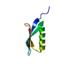


 PDBj
PDBj

 Assembly