 Movie
Movie Controller

[English] 日本語
 Yorodumi
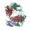
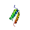
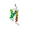
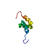
- PDB-1igd: THE THIRD IGG-BINDING DOMAIN FROM STREPTOCOCCAL PROTEIN G: AN ANA... -
+ Open data

- Basic information
| Entry | Database: PDB / ID: 1igd | ||||||
|---|---|---|---|---|---|---|---|
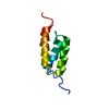
| Title | THE THIRD IGG-BINDING DOMAIN FROM STREPTOCOCCAL PROTEIN G: AN ANALYSIS BY X-RAY CRYSTALLOGRAPHY OF THE STRUCTURE ALONE AND IN A COMPLEX WITH FAB | ||||||
 Components Components | PROTEIN G | ||||||
 Keywords Keywords | IMMUNOGLOBULIN BINDING PROTEIN | ||||||
| Function / homology |  Function and homology information Function and homology information | ||||||
| Biological species |  Streptococcus sp. G148 (bacteria) Streptococcus sp. G148 (bacteria) | ||||||
| Method |  X-RAY DIFFRACTION / Resolution: 1.1 Å X-RAY DIFFRACTION / Resolution: 1.1 Å | ||||||
 Authors Authors | Derrick, J.P. / Wigley, D.B. | ||||||
 Citation Citation | Journal: J.Mol.Biol. / Year: 1994 Title: The third IgG-binding domain from streptococcal protein G. An analysis by X-ray crystallography of the structure alone and in a complex with Fab. Authors: Derrick, J.P. / Wigley, D.B. #1: Journal: Biochemistry / Year: 1994Title: Two Crystal Structures of the B1 Immunoglobulin-Binding Domain of Streptococcal Protein G and Comparison with NMR Authors: Gallagher, T. / Alexander, P. / Bryan, P. / Gilliland, G.L. #2: Journal: Nature / Year: 1992Title: Crystal Structure of a Streptococcal Protein G Domain Bound to an Fab Fragment Authors: Derrick, J.P. / Wigley, D.B. #3: Journal: Biochemistry / Year: 1992Title: 1.67 Angstroms X-Ray Structure of the B2 Immunoglobulin-Binding Domain of Streptococcal Protein G and Comparison to the NMR Structure of the B1 Domain Authors: Achari, A. / Hale, S.P. / Howard, A.J. / Clore, G.M. / Gronenborn, A.M. / Hardman, K.D. / Whitlow, M. #4: Journal: J.Mol.Biol. / Year: 1992Title: Determination of the Solution Structures of Domains II and III of Protein G from Streptococcus by 1H Nuclear Magnetic Resonance Authors: Lian, L.Y. / Derrick, J.P. / Sutcliffe, M.J. / Yang, J.C. / Roberts, G.C.K. #5: Journal: Science / Year: 1991Title: A Novel, Highly Stable Fold of the Immunoglobulin Binding Domain of Streptococcal Protein G Authors: Gronenborn, A.M. / Filpula, D.R. / Essig, N.Z. / Achari, A. / Whitlow, M. / Wingfield, P.T. / Clore, G.M. | ||||||
| History |
|
- Structure visualization
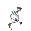
| Structure viewer | Molecule: MolmilJmol/JSmol |
|---|
- Downloads & links
-Download
| PDBx/mmCIF format | 1igd.cif.gz | 25.7 KB | Display | PDBx/mmCIF format |
|---|---|---|---|---|
| PDB format | pdb1igd.ent.gz | 16.5 KB | Display | PDB format |
| PDBx/mmJSON format | 1igd.json.gz | Tree view | PDBx/mmJSON format | |
| Others |  Other downloads Other downloads |
-Validation report
| Arichive directory | https://data.pdbj.org/pub/pdb/validation_reports/ig/1igdftp://data.pdbj.org/pub/pdb/validation_reports/ig/1igd | HTTPS FTP |
|---|
-Related structure data


-Links
 PDBj
PDBj- Assembly
| Deposited unit | 
| ||||||||
|---|---|---|---|---|---|---|---|---|---|
| 1 |
| ||||||||
| Unit cell |
|
-Components
| #1: Protein | Mass: 6657.354 Da / Num. of mol.: 1 Source method: isolated from a genetically manipulated source Source: (gene. exp.) Streptococcus sp. G148 (bacteria) / Plasmid: PUC18 / Species (production host): Escherichia coli / Production host: |
|---|---|
| #2: Water | ChemComp-HOH /  Mass: 18.015 Da / Num. of mol.: 120 / Source method: isolated from a natural source / Formula: H2O Mass: 18.015 Da / Num. of mol.: 120 / Source method: isolated from a natural source / Formula: H2O |
-Experimental details
-Experiment
| Experiment | Method: X-RAY DIFFRACTION |
|---|
- Sample preparation
| Crystal | Density Matthews: 2.23 Å3/Da / Density % sol: 44.81 % | ||||||||||||||||||||||||||||||||||||
|---|---|---|---|---|---|---|---|---|---|---|---|---|---|---|---|---|---|---|---|---|---|---|---|---|---|---|---|---|---|---|---|---|---|---|---|---|---|
| Crystal grow | *PLUS pH: 4.8 / Method: vapor diffusion, hanging drop | ||||||||||||||||||||||||||||||||||||
| Components of the solutions | *PLUS
|
-Data collection
| Radiation | Scattering type: x-ray |
|---|---|
| Radiation wavelength | Relative weight: 1 |
| Reflection | Num. obs: 23530 / % possible obs: 95 % |
| Reflection | *PLUS Highest resolution: 1.1 Å / Lowest resolution: 25 Å / Rmerge(I) obs: 0.058 |
- Processing
| Software |
| ||||||||||||||||||||||||||||||||||||||||||||||||||||||||||||||||||||||||||||||||||||
|---|---|---|---|---|---|---|---|---|---|---|---|---|---|---|---|---|---|---|---|---|---|---|---|---|---|---|---|---|---|---|---|---|---|---|---|---|---|---|---|---|---|---|---|---|---|---|---|---|---|---|---|---|---|---|---|---|---|---|---|---|---|---|---|---|---|---|---|---|---|---|---|---|---|---|---|---|---|---|---|---|---|---|---|---|---|
| Refinement | Resolution: 1.1→25 Å / Rfactor obs: 0.193 | ||||||||||||||||||||||||||||||||||||||||||||||||||||||||||||||||||||||||||||||||||||
| Displacement parameters | Biso mean: 17.7 Å2 | ||||||||||||||||||||||||||||||||||||||||||||||||||||||||||||||||||||||||||||||||||||
| Refine analyze | Luzzati coordinate error obs: 0.05 Å | ||||||||||||||||||||||||||||||||||||||||||||||||||||||||||||||||||||||||||||||||||||
| Refinement step | Cycle: LAST / Resolution: 1.1→25 Å
| ||||||||||||||||||||||||||||||||||||||||||||||||||||||||||||||||||||||||||||||||||||
| Refine LS restraints |
| ||||||||||||||||||||||||||||||||||||||||||||||||||||||||||||||||||||||||||||||||||||
| Software | *PLUS Name: PROLSQ/X-PLOR / Classification: refinement | ||||||||||||||||||||||||||||||||||||||||||||||||||||||||||||||||||||||||||||||||||||
| Refinement | *PLUS Rfactor obs: 0.193 | ||||||||||||||||||||||||||||||||||||||||||||||||||||||||||||||||||||||||||||||||||||
| Solvent computation | *PLUS | ||||||||||||||||||||||||||||||||||||||||||||||||||||||||||||||||||||||||||||||||||||
| Displacement parameters | *PLUS |
