 Movie
Movie Controller

[English] 日本語
 Yorodumi
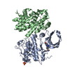
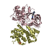
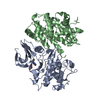
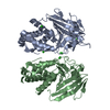
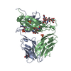
- PDB-1igc: IGG1 FAB FRAGMENT (MOPC21) COMPLEX WITH DOMAIN III OF PROTEIN G F... -
+ Open data

- Basic information
| Entry | Database: PDB / ID: 1igc | ||||||
|---|---|---|---|---|---|---|---|
| Title | IGG1 FAB FRAGMENT (MOPC21) COMPLEX WITH DOMAIN III OF PROTEIN G FROM STREPTOCOCCUS | ||||||
 Components Components |
| ||||||
 Keywords Keywords | COMPLEX (ANTIBODY/BINDING PROTEIN) / PROTEIN G / STREPTOCOCCUS / COMPLEX (ANTIBODY-BINDING PROTEIN) COMPLEX | ||||||
| Function / homology |  Function and homology information Function and homology informationIgG binding / immunoglobulin complex / adaptive immune response / immune response Similarity search - Function | ||||||
| Biological species |   Streptococcus sp. G148 (bacteria) Streptococcus sp. G148 (bacteria) | ||||||
| Method |  X-RAY DIFFRACTION / SYNCHROTRON / Resolution: 2.6 Å X-RAY DIFFRACTION / SYNCHROTRON / Resolution: 2.6 Å | ||||||
 Authors Authors | Derrick, J.P. / Wigley, D.B. | ||||||
 Citation Citation | Journal: J.Mol.Biol. / Year: 1994 Title: The third IgG-binding domain from streptococcal protein G. An analysis by X-ray crystallography of the structure alone and in a complex with Fab. Authors: Derrick, J.P. / Wigley, D.B. #1: Journal: Biochemistry / Year: 1994Title: Two Crystal Structures of the B1 Immunoglobulin-Binding Domain of Streptococcal Protein G and Comparison with NMR Authors: Gallagher, T. / Alexander, P. / Bryan, P. / Gilliland, G.L. #2: Journal: Nature / Year: 1992Title: Crystal Structure of a Streptococcal Protein G Domain Bound to an Fab Fragment Authors: Derrick, J.P. / Wigley, D.B. #3: Journal: J.Mol.Biol. / Year: 1992Title: Crystallization and Preliminary Analysis of the Complex between a Mouse Fab Fragment and a Single Igg-Binding Domain from Streptococcal Protein G Authors: Derrick, J.P. / Davies, G.J. / Dauter, Z. / Wilson, K.S. / Wigley, D.B. #4: Journal: J.Mol.Biol. / Year: 1992Title: Determination of the Solution Structures of Domains II and III of Protein G from Streptococcus by 1H Nuclear Magnetic Resonance Authors: Lian, L.-Y. / Derrick, J.P. / Sutcliff, M.J. / Yang, J.C. / Roberts, G.C.K. #5: Journal: Biochemistry / Year: 1992Title: 1.67 Angstroms X-Ray Structure of the B2 Immunoglobulin-Binding Domain of Streptococcal Protein G and Comparison to the NMR Structure of the B1 Domain Authors: Achari, A. / Hale, S.P. / Howard, A.J. / Clore, G.M. / Gronenborn, A.M. / Hardman, K.D. / Whitlow, M. #6: Journal: Science / Year: 1991Title: A Novel, Highly Stable Fold of the Immunoglobulin Binding Domain of Streptococcal Protein G Authors: Gronenborn, A.M. / Filpula, D.R. / Essig, N.Z. / Achari, A. / Whitlow, M. / Wingfield, P.T. / Clore, G.M. | ||||||
| History |
|
- Structure visualization
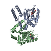
| Structure viewer | Molecule: MolmilJmol/JSmol |
|---|
- Downloads & links
-Download
| PDBx/mmCIF format | 1igc.cif.gz | 120.2 KB | Display | PDBx/mmCIF format |
|---|---|---|---|---|
| PDB format | pdb1igc.ent.gz | 90.6 KB | Display | PDB format |
| PDBx/mmJSON format | 1igc.json.gz | Tree view | PDBx/mmJSON format | |
| Others |  Other downloads Other downloads |
-Validation report
| Arichive directory | https://data.pdbj.org/pub/pdb/validation_reports/ig/1igcftp://data.pdbj.org/pub/pdb/validation_reports/ig/1igc | HTTPS FTP |
|---|
-Related structure data

-Links
 PDBj
PDBj

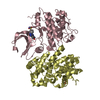
- Assembly
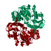
| Deposited unit | 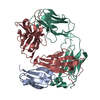
| ||||||||
|---|---|---|---|---|---|---|---|---|---|
| 1 |
| ||||||||
| Unit cell |
| ||||||||
| Atom site foot note | 1: CIS PROLINE - PRO L 8 / 2: CIS PROLINE - PRO L 95 / 3: CIS PROLINE - PRO H 103 / 4: CIS PROLINE - PRO H 154 / 5: CIS PROLINE - PRO H 156 |
-Components
| #1: Antibody | Mass: 23556.004 Da / Num. of mol.: 1 Source method: isolated from a genetically manipulated source Source: (gene. exp.) |
|---|---|
| #2: Antibody | Mass: 23856.750 Da / Num. of mol.: 1 Source method: isolated from a genetically manipulated source Source: (gene. exp.) |
| #3: Protein | Mass: 6657.354 Da / Num. of mol.: 1 Source method: isolated from a genetically manipulated source Source: (gene. exp.) Streptococcus sp. G148 (bacteria) / Plasmid: PUC18 / Production host: |
| #4: Water | ChemComp-HOH /  Mass: 18.015 Da / Num. of mol.: 345 / Source method: isolated from a natural source / Formula: H2O Mass: 18.015 Da / Num. of mol.: 345 / Source method: isolated from a natural source / Formula: H2O |
| Has protein modification | Y |
| Sequence details | THERE ARE NO RESIDUES MISSING FROM THE LIGHT CHAIN. THERE ARE NO RESIDUES MISSING FROM THE N ...THERE ARE NO RESIDUES MISSING FROM THE LIGHT CHAIN. THERE ARE NO RESIDUES MISSING FROM THE N TERMINUS OF THE HEAVY CHAIN. THE SITE OF PROTEOLYSI |
| Source details | MOPC21 (IGG1 KAPPA) WAS ISOLATED FROM ASCITES FLUID, GENERATED BY A MINERAL OIL-INDUCED ...MOPC21 (IGG1 KAPPA) WAS ISOLATED FROM ASCITES FLUID, GENERATED BY A MINERAL OIL-INDUCED PLASMACYTO |
-Experimental details
-Experiment
| Experiment | Method: X-RAY DIFFRACTION |
|---|
- Sample preparation
| Crystal | Density Matthews: 2.52 Å3/Da / Density % sol: 51.26 % | |||||||||||||||||||||||||
|---|---|---|---|---|---|---|---|---|---|---|---|---|---|---|---|---|---|---|---|---|---|---|---|---|---|---|
| Crystal grow | *PLUS pH: 4.8 / Method: vapor diffusion, hanging drop | |||||||||||||||||||||||||
| Components of the solutions | *PLUS
|
-Data collection
| Diffraction source | Source: SYNCHROTRON / Site: EMBL/DESY, HAMBURG  / Wavelength: 1 Å / Wavelength: 1 Å |
|---|---|
| Detector | Date: Apr 1, 1992 |
| Radiation | Monochromatic (M) / Laue (L): M / Scattering type: x-ray |
| Radiation wavelength | Wavelength: 1 Å / Relative weight: 1 |
| Reflection | Num. obs: 17204 / % possible obs: 98.7 % / Rmerge(I) obs: 0.079 |
| Reflection | *PLUS Highest resolution: 2.6 Å / Lowest resolution: 20 Å / Rmerge(I) obs: 0.079 |
- Processing
| Software |
| ||||||||||||||||||||||||||||||||||||||||||||||||||||||||||||||||||||||||||||||||||||
|---|---|---|---|---|---|---|---|---|---|---|---|---|---|---|---|---|---|---|---|---|---|---|---|---|---|---|---|---|---|---|---|---|---|---|---|---|---|---|---|---|---|---|---|---|---|---|---|---|---|---|---|---|---|---|---|---|---|---|---|---|---|---|---|---|---|---|---|---|---|---|---|---|---|---|---|---|---|---|---|---|---|---|---|---|---|
| Refinement | Resolution: 2.6→20 Å / Rfactor obs: 0.168 | ||||||||||||||||||||||||||||||||||||||||||||||||||||||||||||||||||||||||||||||||||||
| Displacement parameters | Biso mean: 31.8 Å2 | ||||||||||||||||||||||||||||||||||||||||||||||||||||||||||||||||||||||||||||||||||||
| Refinement step | Cycle: LAST / Resolution: 2.6→20 Å
| ||||||||||||||||||||||||||||||||||||||||||||||||||||||||||||||||||||||||||||||||||||
| Refine LS restraints |
|
