 Movie
Movie Controller

[English] 日本語
 Yorodumi
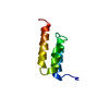
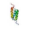
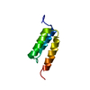
- PDB-2igd: ANISOTROPIC STRUCTURE OF PROTEIN G IGG-BINDING DOMAIN III AT 1.1 ... -
+ Open data

- Basic information
| Entry | Database: PDB / ID: 2igd | ||||||
|---|---|---|---|---|---|---|---|
| Title | ANISOTROPIC STRUCTURE OF PROTEIN G IGG-BINDING DOMAIN III AT 1.1 ANGSTROM RESOLUTION | ||||||
 Components Components | PROTEIN G | ||||||
 Keywords Keywords | IGG-BINDING PROTEIN / ATOMIC RESOLUTION / PROTEIN G / IMMUNOGLOBULIN-BINDING / TRANSMEMBRANE | ||||||
| Function / homology |  Function and homology information Function and homology information | ||||||
| Biological species |  Streptococcus sp. (bacteria) Streptococcus sp. (bacteria) | ||||||
| Method |  X-RAY DIFFRACTION / SYNCHROTRON / INITIAL MODEL WAS PREVIOUSLY OBTAINED STRUCTURE / Resolution: 1.1 Å X-RAY DIFFRACTION / SYNCHROTRON / INITIAL MODEL WAS PREVIOUSLY OBTAINED STRUCTURE / Resolution: 1.1 Å | ||||||
 Authors Authors | Butterworth, S. / Lamzin, V.L. / Wigley, D.B. / Derrick, J.P. / Wilson, K.S. | ||||||
 Citation Citation | Journal: To be Published Title: Anisotropic Refinement of a Protein G Domain at 1.1 Angstrom Resolution Authors: Butterworth, S. / Lamzin, V.S. / Wigley, D.B. / Derrick, J.P. / Wilson, K.S. #1: Journal: J.Mol.Biol. / Year: 1994Title: The Third Igg-Binding Domain from Streptococcal Protein G. An Analysis by X-Ray Crystallography of the Structure Alone and in a Complex with Fab Authors: Derrick, J.P. / Wigley, D.B. | ||||||
| History |
|
- Structure visualization
| Structure viewer | Molecule: MolmilJmol/JSmol |
|---|
- Downloads & links
-Download
| PDBx/mmCIF format | 2igd.cif.gz | 41.1 KB | Display | PDBx/mmCIF format |
|---|---|---|---|---|
| PDB format | pdb2igd.ent.gz | 29.2 KB | Display | PDB format |
| PDBx/mmJSON format | 2igd.json.gz | Tree view | PDBx/mmJSON format | |
| Others |  Other downloads Other downloads |
-Validation report
| Arichive directory | https://data.pdbj.org/pub/pdb/validation_reports/ig/2igdftp://data.pdbj.org/pub/pdb/validation_reports/ig/2igd | HTTPS FTP |
|---|
-Related structure data
| Related structure data | 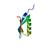 1igdS S: Starting model for refinement |
|---|---|
| Similar structure data |


-Links
 PDBj
PDBj- Assembly
| Deposited unit | 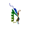
| ||||||||
|---|---|---|---|---|---|---|---|---|---|
| 1 |
| ||||||||
| Unit cell |
|
-Components
| #1: Protein | Mass: 6657.354 Da / Num. of mol.: 1 / Fragment: IMMUNOGLOBULIN-BINDING DOMAIN III Source method: isolated from a genetically manipulated source Source: (gene. exp.) Streptococcus sp. (bacteria) / Strain: G148 / Gene: POTENTIAL / Production host: |
|---|---|
| #2: Water | ChemComp-HOH /  Mass: 18.015 Da / Num. of mol.: 106 / Source method: isolated from a natural source / Formula: H2O Mass: 18.015 Da / Num. of mol.: 106 / Source method: isolated from a natural source / Formula: H2O |
-Experimental details
-Experiment
| Experiment | Method: X-RAY DIFFRACTION / Number of used crystals: 1 |
|---|
- Sample preparation
| Crystal | Density Matthews: 2.23 Å3/Da / Density % sol: 45 % Description: EXTENSION OF FORMER ISOTROPIC MODEL TO ANISOTROPIC MODEL |
|---|---|
| Crystal grow | Method: vapor diffusion, hanging drop / pH: 4.8 Details: CRYSTALS WERE GROWN BY HANGING DROP VAPOUR DIFFUSION FROM 24-26% PEG 4000, 10MM SODIUM ACETATE AT PH 4.8 AND 0.01% SODIUM AZIDE. CELL PARAMETERS ARE NOT THOSE DETERMINED EXPERIMENTALLY. THEY ...Details: CRYSTALS WERE GROWN BY HANGING DROP VAPOUR DIFFUSION FROM 24-26% PEG 4000, 10MM SODIUM ACETATE AT PH 4.8 AND 0.01% SODIUM AZIDE. CELL PARAMETERS ARE NOT THOSE DETERMINED EXPERIMENTALLY. THEY WERE ADJUSTED ON THE BASIS OF THE ENGH & HUBER DICTIONARY AT THE END OF REFINEMENT. THE EXPERIMENTAL ESTIMATES WERE KNOWN TO HAVE POTENTIAL ERRORS DUE TO INACCURACIES IN THE CRYSTAL-TO-DETECTOR AND WAVELENGTH CALIBRATION., vapor diffusion - hanging drop |
-Data collection
| Diffraction | Mean temperature: 300 K |
|---|---|
| Diffraction source | Source: SYNCHROTRON / Site: EMBL/DESY, HAMBURG  / Beamline: X31 / Wavelength: 0.72 / Beamline: X31 / Wavelength: 0.72 |
| Detector | Type: MARRESEARCH / Detector: IMAGE PLATE / Date: Apr 1, 1992 / Details: TOROIDAL MIRROR |
| Radiation | Monochromator: SI(111) / Monochromatic (M) / Laue (L): M / Scattering type: x-ray |
| Radiation wavelength | Wavelength: 0.72 Å / Relative weight: 1 |
| Reflection | Resolution: 1.1→10.1 Å / Num. obs: 24145 / % possible obs: 97.9 % / Observed criterion σ(I): 0 / Redundancy: 4.8 % / Rmerge(I) obs: 0.037 / Rsym value: 0.037 / Net I/σ(I): 39 |
| Reflection shell | Resolution: 1.1→1.12 Å / Redundancy: 3.6 % / Rmerge(I) obs: 0.104 / Mean I/σ(I) obs: 12 / Rsym value: 0.104 / % possible all: 98.1 |
- Processing
| Software |
| |||||||||||||||||||||||||||||||||
|---|---|---|---|---|---|---|---|---|---|---|---|---|---|---|---|---|---|---|---|---|---|---|---|---|---|---|---|---|---|---|---|---|---|---|
| Refinement | Method to determine structure: INITIAL MODEL WAS PREVIOUSLY OBTAINED STRUCTURE Starting model: PDB ENTRY 1IGD Resolution: 1.1→10 Å / Num. parameters: 5485 / Num. restraintsaints: 6708 / Cross valid method: FREE R-VALUE / Stereochemistry target values: ENGH & HUBER Details: PROLSQ AND SHELX-93 ALSO USED IN PRELIMINARY STAGES. SHELX-93 AND SHELX-96 REFINEMENT IS AGAINST INTENSITIES.
| |||||||||||||||||||||||||||||||||
| Solvent computation | Solvent model: SHELX-96 | |||||||||||||||||||||||||||||||||
| Refine analyze | Num. disordered residues: 9 / Occupancy sum hydrogen: 409.9 / Occupancy sum non hydrogen: 571.7 | |||||||||||||||||||||||||||||||||
| Refinement step | Cycle: LAST / Resolution: 1.1→10 Å
| |||||||||||||||||||||||||||||||||
| Refine LS restraints |
|
