back calculated data agree with experimental NOESY spectrum,structures with acceptable covalent geometry,structures with the least restraint violations
Representative
Model #1
closest to the average
-
Components
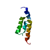
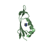
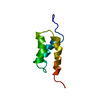
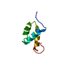
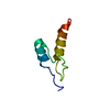
#1: Antibody
ImmunoglobulinGbindingproteinA / IgG binding protein A / Staphylococcal protein A
Mass: 6945.584 Da / Num. of mol.: 1 / Fragment: B DOMAIN / Mutation: Y15W Source method: isolated from a genetically manipulated source Source: (gene. exp.) Staphylococcus aureus (bacteria) / Gene: SPA / Plasmid: pRSET A / Production host: Escherichia coli (E. coli) / Strain (production host): C41 / References: UniProt: P38507
Ionic strength: 100 mM NaCl / 50 mM acetate / pH: 5.5 / Pressure: ambient / Temperature: 298 K
-
NMR measurement
Radiation
Protocol: SINGLE WAVELENGTH / Monochromatic (M) / Laue (L): M
Radiation wavelength
Relative weight: 1
NMR spectrometer
Type: Bruker AMX / Manufacturer: Bruker / Model: AMX / Field strength: 500 MHz
-
Processing
NMR software
Name
Version
Developer
Classification
NMRPipe
2002.044.17.08
Delaglio, F. etal
processing
Sparky
3.106
Goddard, T. D. etal
dataanalysis
CNS
1.1
Brunger, A. T. etal
structuresolution
CNS
1.1
Brunger, A. T. etal
refinement
Refinement
Method: standard anneal.inp CNS script / Software ordinal: 1
NMR representative
Selection criteria: closest to the average
NMR ensemble
Conformer selection criteria: back calculated data agree with experimental NOESY spectrum,structures with acceptable covalent geometry,structures with the least restraint violations Conformers calculated total number: 103 / Conformers submitted total number: 26
+
About Yorodumi
-
News
-
Feb 9, 2022. New format data for meta-information of EMDB entries
New format data for meta-information of EMDB entries
Version 3 of the EMDB header file is now the official format.
The previous official version 1.9 will be removed from the archive.
In the structure databanks used in Yorodumi, some data are registered as the other names, "COVID-19 virus" and "2019-nCoV". Here are the details of the virus and the list of structure data.
Jan 31, 2019. EMDB accession codes are about to change! (news from PDBe EMDB page)
EMDB accession codes are about to change! (news from PDBe EMDB page)
The allocation of 4 digits for EMDB accession codes will soon come to an end. Whilst these codes will remain in use, new EMDB accession codes will include an additional digit and will expand incrementally as the available range of codes is exhausted. The current 4-digit format prefixed with “EMD-” (i.e. EMD-XXXX) will advance to a 5-digit format (i.e. EMD-XXXXX), and so on. It is currently estimated that the 4-digit codes will be depleted around Spring 2019, at which point the 5-digit format will come into force.
The EM Navigator/Yorodumi systems omit the EMD- prefix.
Related info.:Q: What is EMD? / ID/Accession-code notation in Yorodumi/EM Navigator
Yorodumi is a browser for structure data from EMDB, PDB, SASBDB, etc.
This page is also the successor to EM Navigator detail page, and also detail information page/front-end page for Omokage search.
The word "yorodu" (or yorozu) is an old Japanese word meaning "ten thousand". "mi" (miru) is to see.
Related info.:EMDB / PDB / SASBDB / Comparison of 3 databanks / Yorodumi Search / Aug 31, 2016. New EM Navigator & Yorodumi / Yorodumi Papers / Jmol/JSmol / Function and homology information / Changes in new EM Navigator and Yorodumi
 Movie
Movie Controller

 Yorodumi
 Open data
 Basic information
 Components
 Keywords
 Function and homology information
 Staphylococcus aureus (bacteria)
 Authors
 Citation
 Structure visualization
 Downloads & links
 Other downloads
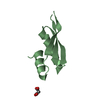


 PDBj
PDBj


 Assembly
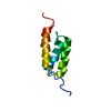
 Sample preparation
 Processing
 NMRPipe