 Movie
Movie Controller

[English] 日本語
 Yorodumi
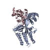
- PDB-6tky: Crystal structure of the DHR2 domain of DOCK10 in complex with CDC42 -
+ Open data

- Basic information
| Entry | Database: PDB / ID: 6tky | ||||||
|---|---|---|---|---|---|---|---|
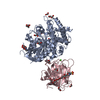
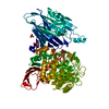
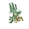
| Title | Crystal structure of the DHR2 domain of DOCK10 in complex with CDC42 | ||||||
 Components Components |
| ||||||
 Keywords Keywords | SIGNALING PROTEIN / DOCK10 GEF CDC42 GTPase | ||||||
| Function / homology |  Function and homology information Function and homology informationmarginal zone B cell differentiation / GBD domain binding / positive regulation of pinocytosis / COG complex / endothelin receptor signaling pathway involved in heart process / storage vacuole / cardiac neural crest cell migration involved in outflow tract morphogenesis / dendritic cell migration / neuron fate determination / apolipoprotein A-I receptor binding ...marginal zone B cell differentiation / GBD domain binding / positive regulation of pinocytosis / COG complex / endothelin receptor signaling pathway involved in heart process / storage vacuole / cardiac neural crest cell migration involved in outflow tract morphogenesis / dendritic cell migration / neuron fate determination / apolipoprotein A-I receptor binding / positive regulation of epithelial cell proliferation involved in lung morphogenesis / regulation of attachment of spindle microtubules to kinetochore / organelle transport along microtubule / Inactivation of CDC42 and RAC1 / positive regulation of pseudopodium assembly / cardiac conduction system development / host-mediated perturbation of viral process / leading edge membrane / regulation of filopodium assembly / neuropilin signaling pathway / establishment of Golgi localization / embryonic heart tube development / dendritic spine morphogenesis / filopodium assembly / cell junction assembly / establishment of epithelial cell apical/basal polarity / adherens junction organization / GTP-dependent protein binding / regulation of Rho protein signal transduction / thioesterase binding / regulation of lamellipodium assembly / regulation of stress fiber assembly / RHO GTPases activate KTN1 / DCC mediated attractive signaling / CD28 dependent Vav1 pathway / regulation of postsynapse organization / positive regulation of filopodium assembly / Wnt signaling pathway, planar cell polarity pathway / phagocytosis, engulfment / RHOV GTPase cycle / Myogenesis / nuclear migration / regulation of mitotic nuclear division / small GTPase-mediated signal transduction / heart contraction / B cell homeostasis / spindle midzone / positive regulation of cytokinesis / establishment of cell polarity / RHOJ GTPase cycle / Golgi organization / RHOQ GTPase cycle / RHOU GTPase cycle / establishment or maintenance of cell polarity / macrophage differentiation / RHO GTPases activate PAKs / CDC42 GTPase cycle / RHOG GTPase cycle / positive regulation of GTPase activity / RAC3 GTPase cycle / regulation of postsynapse assembly / RAC2 GTPase cycle / RHO GTPases Activate WASPs and WAVEs / RHO GTPases activate IQGAPs / negative regulation of protein-containing complex assembly / GPVI-mediated activation cascade / positive regulation of lamellipodium assembly / phagocytic vesicle / positive regulation of stress fiber assembly / RAC1 GTPase cycle / positive regulation of substrate adhesion-dependent cell spreading / EPHB-mediated forward signaling / substantia nigra development / Gene and protein expression by JAK-STAT signaling after Interleukin-12 stimulation / regulation of cell migration / guanyl-nucleotide exchange factor activity / integrin-mediated signaling pathway / actin filament organization / regulation of actin cytoskeleton organization / small monomeric GTPase / FCGR3A-mediated phagocytosis / filopodium / EGFR downregulation / RHO GTPases Activate Formins / Regulation of actin dynamics for phagocytic cup formation / cellular response to type II interferon / VEGFA-VEGFR2 Pathway / MAPK6/MAPK4 signaling / small GTPase binding / endocytosis / apical part of cell / cytoplasmic ribonucleoprotein granule / cell-cell junction / mitotic spindle / G beta:gamma signalling through CDC42 / intracellular protein localization / ubiquitin protein ligase activity / Factors involved in megakaryocyte development and platelet production / positive regulation of cell growth / actin cytoskeleton organization Similarity search - Function | ||||||
| Biological species |  Homo sapiens (human) Homo sapiens (human) | ||||||
| Method |  X-RAY DIFFRACTION / SYNCHROTRON / MOLECULAR REPLACEMENT / Resolution: 2.55 Å X-RAY DIFFRACTION / SYNCHROTRON / MOLECULAR REPLACEMENT / Resolution: 2.55 Å | ||||||
 Authors Authors | Barford, D. / Fan, D. / Cronin, N. / Yang, J. | ||||||
| Funding support |  United Kingdom, 1items United Kingdom, 1items
| ||||||
 Citation Citation | Journal: Biorxiv / Year: 2022 Title: Structural basis for CDC42 and RAC activation by the dual specificity GEF DOCK10 Authors: Fan, D. / Yang, J. / Cronin, N. / Barford, D. | ||||||
| History |
|
- Structure visualization
| Structure viewer | Molecule: MolmilJmol/JSmol |
|---|
- Downloads & links
-Download
| PDBx/mmCIF format | 6tky.cif.gz | 308.2 KB | Display | PDBx/mmCIF format |
|---|---|---|---|---|
| PDB format | pdb6tky.ent.gz | 195.2 KB | Display | PDB format |
| PDBx/mmJSON format | 6tky.json.gz | Tree view | PDBx/mmJSON format | |
| Others |  Other downloads Other downloads |
-Validation report
| Arichive directory | https://data.pdbj.org/pub/pdb/validation_reports/tk/6tkyftp://data.pdbj.org/pub/pdb/validation_reports/tk/6tky | HTTPS FTP |
|---|
-Related structure data
| Related structure data |  6tkzC 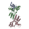 6tm1C 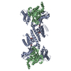 2wm9S S: Starting model for refinement C: citing same article ( |
|---|---|
| Similar structure data |


-Links
 PDBj
PDBj


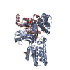
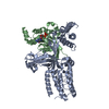
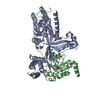
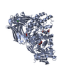
- Assembly
| Deposited unit | 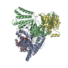
| ||||||||||||
|---|---|---|---|---|---|---|---|---|---|---|---|---|---|
| 1 | 
| ||||||||||||
| 2 | 
| ||||||||||||
| Unit cell |
|
-Components
| #1: Protein | Mass: 53026.414 Da / Num. of mol.: 1 Source method: isolated from a genetically manipulated source Details: NA / Source: (gene. exp.) Homo sapiens (human) / Tissue: NA / Cell: NA / Cell line: NA / Gene: DOCK10, KIAA0694, ZIZ3 / Organ: NA / Variant: NA / Production host:  | ||||||||
|---|---|---|---|---|---|---|---|---|---|
| #2: Protein | Mass: 20958.135 Da / Num. of mol.: 2 Source method: isolated from a genetically manipulated source Source: (gene. exp.) Homo sapiens (human) / Gene: CDC42 / Production host: #3: Protein | | Mass: 53083.465 Da / Num. of mol.: 1 Source method: isolated from a genetically manipulated source Source: (gene. exp.) Homo sapiens (human) / Gene: DOCK10, KIAA0694, ZIZ3 / Production host: #4: Chemical |   Mass: 92.094 Da / Num. of mol.: 3 / Source method: isolated from a natural source / Formula: C3H8O3 / Feature type: SUBJECT OF INVESTIGATION Mass: 92.094 Da / Num. of mol.: 3 / Source method: isolated from a natural source / Formula: C3H8O3 / Feature type: SUBJECT OF INVESTIGATION#5: Water | ChemComp-HOH / |  Mass: 18.015 Da / Num. of mol.: 231 / Source method: isolated from a natural source / Formula: H2O Mass: 18.015 Da / Num. of mol.: 231 / Source method: isolated from a natural source / Formula: H2OHas ligand of interest | Y | |
-Experimental details
-Experiment
| Experiment | Method: X-RAY DIFFRACTION / Number of used crystals: 1 |
|---|
- Sample preparation
| Crystal | Density Matthews: 2.48 Å3/Da / Density % sol: 50.5 % |
|---|---|
| Crystal grow | Temperature: 293 K / Method: evaporation / pH: 7.5 Details: 25% (w/v) PEG 3350, 200 mM potassium acetate, 8% (v/v) 1,1,1,3,3,3-Hexafluoro-2-propanol. |
-Data collection
| Diffraction | Mean temperature: 100 K / Serial crystal experiment: N |
|---|---|
| Diffraction source | Source: SYNCHROTRON / Site: Diamond / Beamline: I02 / Wavelength: 0.979 Å |
| Detector | Type: DECTRIS PILATUS3 2M / Detector: PIXEL / Date: Sep 9, 2010 |
| Radiation | Protocol: SINGLE WAVELENGTH / Monochromatic (M) / Laue (L): M / Scattering type: x-ray |
| Radiation wavelength | Wavelength: 0.979 Å / Relative weight: 1 |
| Reflection | Resolution: 2.55→29.83 Å / Num. obs: 49296 / % possible obs: 99.9 % / Redundancy: 7.23 % / Biso Wilson estimate: 42.35 Å2 / Rsym value: 0.126 / Net I/σ(I): 5.52 |
| Reflection shell | Resolution: 2.55→2.64 Å / Num. unique obs: 5918 / Rsym value: 0.34 |
- Processing
| Software |
| |||||||||||||||||||||||||||||||||||||||||||||||||||||||||||||||||||||||||||||||||||||||||||||||||||||||||||||||||||||||||||||||||||||
|---|---|---|---|---|---|---|---|---|---|---|---|---|---|---|---|---|---|---|---|---|---|---|---|---|---|---|---|---|---|---|---|---|---|---|---|---|---|---|---|---|---|---|---|---|---|---|---|---|---|---|---|---|---|---|---|---|---|---|---|---|---|---|---|---|---|---|---|---|---|---|---|---|---|---|---|---|---|---|---|---|---|---|---|---|---|---|---|---|---|---|---|---|---|---|---|---|---|---|---|---|---|---|---|---|---|---|---|---|---|---|---|---|---|---|---|---|---|---|---|---|---|---|---|---|---|---|---|---|---|---|---|---|---|---|
| Refinement | Method to determine structure: MOLECULAR REPLACEMENT Starting model: 2WM9 Resolution: 2.55→29.83 Å / SU ML: 0.3249 / Cross valid method: FREE R-VALUE / σ(F): 1.35 / Phase error: 23.18
| |||||||||||||||||||||||||||||||||||||||||||||||||||||||||||||||||||||||||||||||||||||||||||||||||||||||||||||||||||||||||||||||||||||
| Solvent computation | Shrinkage radii: 0.9 Å / VDW probe radii: 1.11 Å | |||||||||||||||||||||||||||||||||||||||||||||||||||||||||||||||||||||||||||||||||||||||||||||||||||||||||||||||||||||||||||||||||||||
| Displacement parameters | Biso mean: 45.26 Å2 | |||||||||||||||||||||||||||||||||||||||||||||||||||||||||||||||||||||||||||||||||||||||||||||||||||||||||||||||||||||||||||||||||||||
| Refinement step | Cycle: LAST / Resolution: 2.55→29.83 Å
| |||||||||||||||||||||||||||||||||||||||||||||||||||||||||||||||||||||||||||||||||||||||||||||||||||||||||||||||||||||||||||||||||||||
| Refine LS restraints |
| |||||||||||||||||||||||||||||||||||||||||||||||||||||||||||||||||||||||||||||||||||||||||||||||||||||||||||||||||||||||||||||||||||||
| LS refinement shell |
|
