 Movie
Movie Controller

[English] 日本語
 Yorodumi
- PDB-6tm1: Crystal structure of the DHR2 domain of DOCK10 in complex with RAC3 -
+ Open data

- Basic information
| Entry | Database: PDB / ID: 6tm1 | ||||||
|---|---|---|---|---|---|---|---|
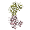
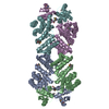
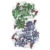
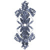
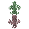
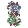
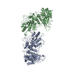
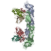
| Title | Crystal structure of the DHR2 domain of DOCK10 in complex with RAC3 | ||||||
 Components Components |
| ||||||
 Keywords Keywords | SIGNALING PROTEIN / DOCK10 GEF RAC3 GTPase | ||||||
| Function / homology |  Function and homology information Function and homology informationmarginal zone B cell differentiation / postsynaptic actin cytoskeleton organization / cerebral cortex GABAergic interneuron development / regulation of neutrophil migration / NADPH oxidase complex / regulation of neuron maturation / respiratory burst / cortical cytoskeleton organization / dendritic spine morphogenesis / regulation of Rho protein signal transduction ...marginal zone B cell differentiation / postsynaptic actin cytoskeleton organization / cerebral cortex GABAergic interneuron development / regulation of neutrophil migration / NADPH oxidase complex / regulation of neuron maturation / respiratory burst / cortical cytoskeleton organization / dendritic spine morphogenesis / regulation of Rho protein signal transduction / cell projection assembly / motor neuron axon guidance / PCP/CE pathway / positive regulation of cell adhesion mediated by integrin / neuromuscular process controlling balance / small GTPase-mediated signal transduction / synaptic transmission, GABAergic / B cell homeostasis / filamentous actin / establishment or maintenance of cell polarity / Rac protein signal transduction / CDC42 GTPase cycle / positive regulation of GTPase activity / RAC3 GTPase cycle / regulation of postsynapse assembly / RAC2 GTPase cycle / RAC1 GTPase cycle / positive regulation of substrate adhesion-dependent cell spreading / endomembrane system / regulation of cell migration / homeostasis of number of cells within a tissue / guanyl-nucleotide exchange factor activity / cell periphery / actin filament organization / small monomeric GTPase / regulation of actin cytoskeleton organization / cell chemotaxis / small GTPase binding / Wnt signaling pathway / calcium-dependent protein binding / regulation of cell shape / lamellipodium / Factors involved in megakaryocyte development and platelet production / growth cone / actin cytoskeleton organization / G protein activity / cytoplasmic vesicle / dendritic spine / cytoskeleton / neuron projection / intracellular signal transduction / postsynapse / neuronal cell body / GTPase activity / protein kinase binding / endoplasmic reticulum membrane / GTP binding / perinuclear region of cytoplasm / glutamatergic synapse / extracellular exosome / nucleoplasm / membrane / nucleus / plasma membrane / cytoplasm / cytosol Similarity search - Function | ||||||
| Biological species |  Homo sapiens (human) Homo sapiens (human) | ||||||
| Method |  X-RAY DIFFRACTION / SYNCHROTRON / MOLECULAR REPLACEMENT / Resolution: 3.71 Å X-RAY DIFFRACTION / SYNCHROTRON / MOLECULAR REPLACEMENT / Resolution: 3.71 Å | ||||||
 Authors Authors | Barford, D. / Fan, D. / Cronin, N. / Yang, J. | ||||||
| Funding support |  United Kingdom, 1items United Kingdom, 1items
| ||||||
 Citation Citation | Journal: Biorxiv / Year: 2022 Title: Structural basis for CDC42 and RAC activation by the dual specificity GEF DOCK10 Authors: Fan, D. / Yang, J. / Cronin, N. / Barford, D. | ||||||
| History |
|
- Structure visualization
| Structure viewer | Molecule: MolmilJmol/JSmol |
|---|
- Downloads & links
-Download
| PDBx/mmCIF format | 6tm1.cif.gz | 234.1 KB | Display | PDBx/mmCIF format |
|---|---|---|---|---|
| PDB format | pdb6tm1.ent.gz | 144.3 KB | Display | PDB format |
| PDBx/mmJSON format | 6tm1.json.gz | Tree view | PDBx/mmJSON format | |
| Others |  Other downloads Other downloads |
-Validation report
| Arichive directory | https://data.pdbj.org/pub/pdb/validation_reports/tm/6tm1ftp://data.pdbj.org/pub/pdb/validation_reports/tm/6tm1 | HTTPS FTP |
|---|
-Related structure data
| Related structure data | 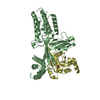 6tkyC 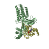 6tkzC 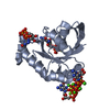 2c2hS 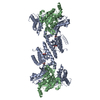 2wm9S S: Starting model for refinement C: citing same article ( |
|---|---|
| Similar structure data |
-Links
 PDBj
PDBj


- Assembly
| Deposited unit | 
| ||||||||||||
|---|---|---|---|---|---|---|---|---|---|---|---|---|---|
| 1 | 
| ||||||||||||
| Unit cell |
|
-Components
| #1: Protein | Mass: 53026.414 Da / Num. of mol.: 1 Source method: isolated from a genetically manipulated source Details: ...Details: STPELRRTWLESMAKIHARNGDLSEAAMCYIHIAALIAEYLKRKGYWKVEKICTASLLSEDTHPCDSNSLLTTPSGGSMFSMGWPAFLSITPNIKEEGAMKEDSGMQDTPYNENILVEQLYMCVEFLWKSERYELIADVNKPIIAVFEKQRDFKKLSDLYYDIHRSYLKVAEVVNSEKRLFGRYYRVAFYGQGFFEE EEGKEYIYKEPKLTGLSEISQRLLKLYADKFGADNVKIIQDSNKVNPKDLDPKYAYIQVTYVTPFFEEKEIEDRKTDFEMHHNINRFVFETPFTLSGKKH GGVAEQCKRRTILTTSHLFPYVKKRIQVISQSSTELNPIEVAIDEMSKKVSELNQLCTMEEVDMIRLQLKLQGSVSVKVNAGPMAYARAFLEETNAKKYP DNQVKLLKEIFRQFADACGQ ALDVNERLIK EDQLEYQEEL RSHYKDMLSE LSTVMNEQIT Source: (gene. exp.) Homo sapiens (human) / Gene: DOCK10, KIAA0694, ZIZ3 / Production host:  |
|---|---|
| #2: Protein | Mass: 21405.861 Da / Num. of mol.: 1 Source method: isolated from a genetically manipulated source Source: (gene. exp.) Homo sapiens (human) / Gene: RAC3 / Production host: |
| #3: Protein | Mass: 53083.465 Da / Num. of mol.: 1 Source method: isolated from a genetically manipulated source Source: (gene. exp.) Homo sapiens (human) / Gene: DOCK10, KIAA0694, ZIZ3 / Production host: |
-Experimental details
-Experiment
| Experiment | Method: X-RAY DIFFRACTION / Number of used crystals: 1 |
|---|
- Sample preparation
| Crystal | Density Matthews: 2.98 Å3/Da / Density % sol: 58.75 % |
|---|---|
| Crystal grow | Temperature: 293 K / Method: evaporation / pH: 7.5 Details: 25% (w/v) PEG 1500, 100 mM MMT. MMT buffer is produced by mixing DL-malic acid, MES and Tris base in the molar ratios of 1:2:2. |
-Data collection
| Diffraction | Mean temperature: 100 K / Serial crystal experiment: N |
|---|---|
| Diffraction source | Source: SYNCHROTRON / Site: Diamond / Beamline: I04 / Wavelength: 0.98 Å |
| Detector | Type: DECTRIS PILATUS 2M / Detector: PIXEL / Date: Sep 9, 2011 |
| Radiation | Protocol: SINGLE WAVELENGTH / Monochromatic (M) / Laue (L): M / Scattering type: x-ray |
| Radiation wavelength | Wavelength: 0.98 Å / Relative weight: 1 |
| Reflection | Resolution: 3.71→29.6 Å / Num. obs: 29674 / % possible obs: 95.5 % / Redundancy: 2.6 % / Biso Wilson estimate: 151.85 Å2 / Rmerge(I) obs: 0.08 / Net I/σ(I): 6.61 |
| Reflection shell | Resolution: 3.71→3.85 Å / Rmerge(I) obs: 0.3 / Num. unique obs: 1515 |
- Processing
| Software |
| ||||||||||||||||||||||||||||||||||||||||||||||||||||||||||||||||||||||||||||||||||||
|---|---|---|---|---|---|---|---|---|---|---|---|---|---|---|---|---|---|---|---|---|---|---|---|---|---|---|---|---|---|---|---|---|---|---|---|---|---|---|---|---|---|---|---|---|---|---|---|---|---|---|---|---|---|---|---|---|---|---|---|---|---|---|---|---|---|---|---|---|---|---|---|---|---|---|---|---|---|---|---|---|---|---|---|---|---|
| Refinement | Method to determine structure: MOLECULAR REPLACEMENT Starting model: 2WM9,2C2H Resolution: 3.71→29.6 Å / SU ML: 0.6511 / Cross valid method: FREE R-VALUE / σ(F): 1.18 / Phase error: 38.1002 Stereochemistry target values: GeoStd + Monomer Library + CDL v1.2
| ||||||||||||||||||||||||||||||||||||||||||||||||||||||||||||||||||||||||||||||||||||
| Solvent computation | Shrinkage radii: 0.9 Å / VDW probe radii: 1.11 Å / Solvent model: FLAT BULK SOLVENT MODEL | ||||||||||||||||||||||||||||||||||||||||||||||||||||||||||||||||||||||||||||||||||||
| Displacement parameters | Biso mean: 148.82 Å2 | ||||||||||||||||||||||||||||||||||||||||||||||||||||||||||||||||||||||||||||||||||||
| Refinement step | Cycle: LAST / Resolution: 3.71→29.6 Å
| ||||||||||||||||||||||||||||||||||||||||||||||||||||||||||||||||||||||||||||||||||||
| Refine LS restraints |
| ||||||||||||||||||||||||||||||||||||||||||||||||||||||||||||||||||||||||||||||||||||
| LS refinement shell |
|
