 Movie
Movie Controller

[English] 日本語
 Yorodumi
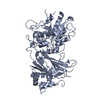
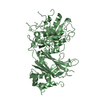
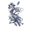
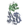
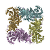
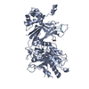
- PDB-1qat: 1-PHOSPHATIDYLINOSITOL-4,5-BISPHOSPHATE PHOSPHODIESTERASE DELTA C... -
+ Open data

- Basic information
| Entry | Database: PDB / ID: 1qat | ||||||
|---|---|---|---|---|---|---|---|
| Title | 1-PHOSPHATIDYLINOSITOL-4,5-BISPHOSPHATE PHOSPHODIESTERASE DELTA COMPLEX WITH SAMARIUM (III) CHLORIDE | ||||||
 Components Components | PHOSPHOLIPASE C DELTA-1 | ||||||
 Keywords Keywords | HYDROLASE / LIPID DEGRADATION / TRANSDUCER / CALCIUM-BINDING | ||||||
| Function / homology |  Function and homology information Function and homology informationphospholipase C/protein kinase C signal transduction / positive regulation of inositol trisphosphate biosynthetic process / Synthesis of IP3 and IP4 in the cytosol / response to prostaglandin F / phosphatidylinositol phospholipase C activity / phosphoinositide phospholipase C / phosphatidylinositol phosphate binding / response to aluminum ion / positive regulation of norepinephrine secretion / phosphatidylinositol-4,5-bisphosphate 5-phosphatase activity ...phospholipase C/protein kinase C signal transduction / positive regulation of inositol trisphosphate biosynthetic process / Synthesis of IP3 and IP4 in the cytosol / response to prostaglandin F / phosphatidylinositol phospholipase C activity / phosphoinositide phospholipase C / phosphatidylinositol phosphate binding / response to aluminum ion / positive regulation of norepinephrine secretion / phosphatidylinositol-4,5-bisphosphate 5-phosphatase activity / phosphatidylinositol metabolic process / phosphatidylinositol-4,5-bisphosphate phospholipase C activity / C-type glycerophospholipase activity / calcium-dependent phospholipid binding / phosphatidic acid binding / inositol 1,4,5 trisphosphate binding / GTPase activating protein binding / labyrinthine layer blood vessel development / response to hyperoxia / lipid catabolic process / phosphatidylinositol-4,5-bisphosphate binding / cellular response to calcium ion / phospholipid binding / response to calcium ion / response to peptide hormone / mitochondrial membrane / regulation of cell population proliferation / angiogenesis / phospholipase C-activating G protein-coupled receptor signaling pathway / G protein-coupled receptor signaling pathway / calcium ion binding / enzyme binding / plasma membrane / cytosol / cytoplasm Similarity search - Function | ||||||
| Biological species |  | ||||||
| Method |  X-RAY DIFFRACTION / DIFFERENCE FOURIER / Resolution: 3 Å X-RAY DIFFRACTION / DIFFERENCE FOURIER / Resolution: 3 Å | ||||||
 Authors Authors | Grobler, J.A. / Hurley, J.H. | ||||||
 Citation Citation | Journal: Nat.Struct.Biol. / Year: 1996 Title: C2 domain conformational changes in phospholipase C-delta 1. Authors: Grobler, J.A. / Essen, L.O. / Williams, R.L. / Hurley, J.H. #1: Journal: Nature / Year: 1996Title: Crystal Structure of a Mammalian Phosphoinositide-Specific Phospholipase C Delta Authors: Essen, L.O. / Perisic, O. / Cheung, R. / Katan, M. / Williams, R.L. #2: Journal: Protein Sci. / Year: 1996Title: Expression, Characterization, and Crystallization of the Catalytic Core of Rat Phosphatidylinositide-Specific Phospholipase C Delta 1 Authors: Grobler, J.A. / Hurley, J.H. | ||||||
| History |
|
- Structure visualization
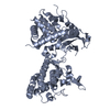
| Structure viewer | Molecule: MolmilJmol/JSmol |
|---|
- Downloads & links
-Download
| PDBx/mmCIF format | 1qat.cif.gz | 209.9 KB | Display | PDBx/mmCIF format |
|---|---|---|---|---|
| PDB format | pdb1qat.ent.gz | 164.7 KB | Display | PDB format |
| PDBx/mmJSON format | 1qat.json.gz | Tree view | PDBx/mmJSON format | |
| Others |  Other downloads Other downloads |
-Validation report
| Arichive directory | https://data.pdbj.org/pub/pdb/validation_reports/qa/1qatftp://data.pdbj.org/pub/pdb/validation_reports/qa/1qat | HTTPS FTP |
|---|
-Related structure data

-Links
 PDBj
PDBj


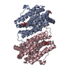
- Assembly
| Deposited unit | 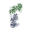
| ||||||||
|---|---|---|---|---|---|---|---|---|---|
| 1 | 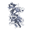
| ||||||||
| 2 | 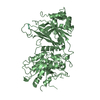
| ||||||||
| Unit cell |
|
-Components
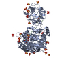
| #1: Protein | Mass: 70430.383 Da / Num. of mol.: 2 Source method: isolated from a genetically manipulated source Details: PHOSHOINOSITIDE-SPECIFIC PHOSPHOLIPASE C DELTA-1 / Source: (gene. exp.)  References: UniProt: P10688, phosphoinositide phospholipase C #2: Chemical | ChemComp-SM /   Mass: 150.360 Da / Num. of mol.: 5 / Source method: obtained synthetically / Formula: Sm Mass: 150.360 Da / Num. of mol.: 5 / Source method: obtained synthetically / Formula: Sm |
|---|
-Experimental details
-Experiment
| Experiment | Method: X-RAY DIFFRACTION / Number of used crystals: 1 |
|---|
- Sample preparation
| Crystal | Density Matthews: 2.59 Å3/Da / Density % sol: 52.51 % | ||||||||||||||||||||||||||||||
|---|---|---|---|---|---|---|---|---|---|---|---|---|---|---|---|---|---|---|---|---|---|---|---|---|---|---|---|---|---|---|---|
| Crystal grow | Method: clusters formed by mixing - used as seeds in hanging drop pH: 6.5 Details: NEEDLE CLUSTERS WERE FORMED BY MIXING EQUAL VOLUMES OF PROTEIN SOLUTION (22 MG/ML) WITH A WELL SOLUTION CONSISTING OF 0.1 M NA MES (PH 6.0), 0.2 M LICL, 20% GLYCEROL, AND 12-14 % PEG 8000. ...Details: NEEDLE CLUSTERS WERE FORMED BY MIXING EQUAL VOLUMES OF PROTEIN SOLUTION (22 MG/ML) WITH A WELL SOLUTION CONSISTING OF 0.1 M NA MES (PH 6.0), 0.2 M LICL, 20% GLYCEROL, AND 12-14 % PEG 8000. FRAGMENTS OF THE NEEDLE CLUSTERS WERE USED TO SEED HANGING DROPS. THE WELL SOLUTION USED FOR THE SEEDING EXPERIMENTS WAS ADJUSTED TO 0.1M NA MES (PH 6.5), 0.2 M LICL, 20 % GLYCEROL, AND 6-8 % PEG 8000., clusters formed by mixing - used as seeds in hanging drops PH range: 6.0-6.5 | ||||||||||||||||||||||||||||||
| Crystal grow | *PLUS Method: vapor diffusion, hanging dropDetails: used to seeding, Grobler, J.A., (1996) Protein Sci., 5, 680. | ||||||||||||||||||||||||||||||
| Components of the solutions | *PLUS
|
-Data collection
| Diffraction | Mean temperature: 100 K |
|---|---|
| Diffraction source | Source: ROTATING ANODE / Type: RIGAKU RUH2R / Wavelength: 1.5418 |
| Detector | Type: RIGAKU RAXIS IIC / Detector: IMAGE PLATE / Details: MIRRORS |
| Radiation | Monochromatic (M) / Laue (L): M / Scattering type: x-ray |
| Radiation wavelength | Wavelength: 1.5418 Å / Relative weight: 1 |
| Reflection | Resolution: 3→60 Å / Num. obs: 24235 / % possible obs: 84.9 % / Observed criterion σ(I): -2 / Redundancy: 2.5 % / Rmerge(I) obs: 0.121 / Rsym value: 0.121 / Net I/σ(I): 7.2 |
| Reflection shell | Resolution: 3→3.11 Å / Redundancy: 1.8 % / Rmerge(I) obs: 0.248 / Mean I/σ(I) obs: 2.3 / Rsym value: 0.248 / % possible all: 73.8 |
- Processing
| Software |
| ||||||||||||||||||||||||||||||||||||||||||||||||||||||||||||
|---|---|---|---|---|---|---|---|---|---|---|---|---|---|---|---|---|---|---|---|---|---|---|---|---|---|---|---|---|---|---|---|---|---|---|---|---|---|---|---|---|---|---|---|---|---|---|---|---|---|---|---|---|---|---|---|---|---|---|---|---|---|
| Refinement | Method to determine structure: DIFFERENCE FOURIER Starting model: APO TRICLINIC PHOSPHOLIPASE C Resolution: 3→6 Å / Cross valid method: FREE R
| ||||||||||||||||||||||||||||||||||||||||||||||||||||||||||||
| Refine analyze |
| ||||||||||||||||||||||||||||||||||||||||||||||||||||||||||||
| Refinement step | Cycle: LAST / Resolution: 3→6 Å
| ||||||||||||||||||||||||||||||||||||||||||||||||||||||||||||
| Refine LS restraints |
| ||||||||||||||||||||||||||||||||||||||||||||||||||||||||||||
| LS refinement shell | Resolution: 3→3.09 Å /
| ||||||||||||||||||||||||||||||||||||||||||||||||||||||||||||
| Software | *PLUS Name: X-PLOR / Classification: refinement | ||||||||||||||||||||||||||||||||||||||||||||||||||||||||||||
| Refine LS restraints | *PLUS
|
