 Movie
Movie Controller

[English] 日本語
 Yorodumi
- PDB-1uzr: Crystal Structure of the Class Ib Ribonucleotide Reductase R2F-2 ... -
+ Open data

- Basic information
| Entry | Database: PDB / ID: 1uzr | ||||||
|---|---|---|---|---|---|---|---|
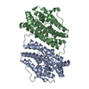
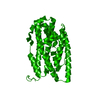
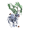
| Title | Crystal Structure of the Class Ib Ribonucleotide Reductase R2F-2 subunit from Mycobacterium tuberculosis | ||||||
 Components Components | RIBONUCLEOTIDE REDUCTASE R2-2 SMALL SUBUNIT | ||||||
 Keywords Keywords | REDUCTASE / RIBONUCLEOTIDE REDUCTASE / TUBERCULOSIS / R2F-2 / RADICAL GENERATION / SMALL SUBUNIT | ||||||
| Function / homology |  Function and homology information Function and homology informationribonucleoside-diphosphate reductase complex / ribonucleoside-diphosphate reductase / ribonucleoside-diphosphate reductase activity, thioredoxin disulfide as acceptor / deoxyribonucleotide biosynthetic process / DNA replication / metal ion binding Similarity search - Function | ||||||
| Biological species |   MYCOBACTERIUM TUBERCULOSIS (bacteria) MYCOBACTERIUM TUBERCULOSIS (bacteria) | ||||||
| Method |  X-RAY DIFFRACTION / SYNCHROTRON / MOLECULAR REPLACEMENT / Resolution: 2.2 Å X-RAY DIFFRACTION / SYNCHROTRON / MOLECULAR REPLACEMENT / Resolution: 2.2 Å | ||||||
 Authors Authors | Uppsten, M. / Davis, J. / Rubin, H. / Uhlin, U. | ||||||
 Citation Citation | Journal: FEBS Lett. / Year: 2004 Title: Crystal Structure of the Biologically Active Form of Class Ib Ribonucleotide Reductase Small Subunit from Mycobacterium Tuberculosis Authors: Uppsten, M. / Davis, J. / Rubin, H. / Uhlin, U. | ||||||
| History |
|
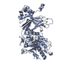
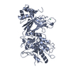
- Structure visualization
| Structure viewer | Molecule: MolmilJmol/JSmol |
|---|
- Downloads & links
-Download
| PDBx/mmCIF format | 1uzr.cif.gz | 189.9 KB | Display | PDBx/mmCIF format |
|---|---|---|---|---|
| PDB format | pdb1uzr.ent.gz | 152.2 KB | Display | PDB format |
| PDBx/mmJSON format | 1uzr.json.gz | Tree view | PDBx/mmJSON format | |
| Others |  Other downloads Other downloads |
-Validation report
| Arichive directory | https://data.pdbj.org/pub/pdb/validation_reports/uz/1uzrftp://data.pdbj.org/pub/pdb/validation_reports/uz/1uzr | HTTPS FTP |
|---|
-Related structure data
| Related structure data |  1r2fS S: Starting model for refinement |
|---|---|
| Similar structure data |


-Links
 PDBj
PDBj
- Assembly
| Deposited unit | 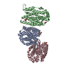
| ||||||||||||
|---|---|---|---|---|---|---|---|---|---|---|---|---|---|
| 1 | 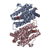
| ||||||||||||
| 2 | 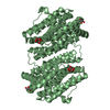
| ||||||||||||
| Unit cell |
| ||||||||||||
| Noncrystallographic symmetry (NCS) | NCS oper:
|
-Components
| #1: Protein | Mass: 33842.078 Da / Num. of mol.: 3 / Fragment: RADICAL GENERATING SUBUNIT, RESIDUES 1-296 Source method: isolated from a genetically manipulated source Source: (gene. exp.) MYCOBACTERIUM TUBERCULOSIS (bacteria) / Production host: References: UniProt: Q50549, UniProt: P9WH71*PLUS, ribonucleoside-diphosphate reductase #2: Chemical | ChemComp-FE /   Mass: 55.845 Da / Num. of mol.: 6 / Source method: obtained synthetically / Formula: Fe Mass: 55.845 Da / Num. of mol.: 6 / Source method: obtained synthetically / Formula: Fe#3: Chemical |   Mass: 192.124 Da / Num. of mol.: 3 / Source method: obtained synthetically / Formula: C6H8O7 Mass: 192.124 Da / Num. of mol.: 3 / Source method: obtained synthetically / Formula: C6H8O7#4: Chemical |   Mass: 92.094 Da / Num. of mol.: 3 / Source method: obtained synthetically / Formula: C3H8O3 Mass: 92.094 Da / Num. of mol.: 3 / Source method: obtained synthetically / Formula: C3H8O3#5: Water | ChemComp-HOH / |  Mass: 18.015 Da / Num. of mol.: 391 / Source method: isolated from a natural source / Formula: H2O Mass: 18.015 Da / Num. of mol.: 391 / Source method: isolated from a natural source / Formula: H2O |
|---|
-Experimental details
-Experiment
| Experiment | Method: X-RAY DIFFRACTION / Number of used crystals: 1 |
|---|
- Sample preparation
| Crystal | Density Matthews: 3.4 Å3/Da / Density % sol: 63.3 % |
|---|---|
| Crystal grow | pH: 5.6 / Details: 0.6 M AMSO4, 100MM NACT PH 5.6, 50MM NA/K TARTRATE |
-Data collection
| Diffraction | Mean temperature: 100 K |
|---|---|
| Diffraction source | Source: SYNCHROTRON / Site: ESRF  / Beamline: ID29 / Wavelength: 0.976 / Beamline: ID29 / Wavelength: 0.976 |
| Detector | Type: ADSC CCD / Detector: CCD / Date: Aug 15, 2003 |
| Radiation | Monochromator: CRYOGENICALLY COOLED HIGH RESOLUTION SI (311) AND LOWER RESOLUTION SI (111) Protocol: SINGLE WAVELENGTH / Monochromatic (M) / Laue (L): M / Scattering type: x-ray |
| Radiation wavelength | Wavelength: 0.976 Å / Relative weight: 1 |
| Reflection | Resolution: 2.2→40 Å / Num. obs: 77125 / % possible obs: 99 % / Redundancy: 4.84 % / Rmerge(I) obs: 0.091 / Net I/σ(I): 10 |
| Reflection shell | Resolution: 2.2→2.25 Å / Rmerge(I) obs: 0.445 / Mean I/σ(I) obs: 1.7 / % possible all: 93.5 |
- Processing
| Software |
| ||||||||||||||||||||||||||||||||||||||||||||||||||||||||||||||||||||||||||||||||||||||||||||||||||||||||||||||||||||||||||||||||||||||||||||||||||||||||||||||||||||||||||||||||||||||
|---|---|---|---|---|---|---|---|---|---|---|---|---|---|---|---|---|---|---|---|---|---|---|---|---|---|---|---|---|---|---|---|---|---|---|---|---|---|---|---|---|---|---|---|---|---|---|---|---|---|---|---|---|---|---|---|---|---|---|---|---|---|---|---|---|---|---|---|---|---|---|---|---|---|---|---|---|---|---|---|---|---|---|---|---|---|---|---|---|---|---|---|---|---|---|---|---|---|---|---|---|---|---|---|---|---|---|---|---|---|---|---|---|---|---|---|---|---|---|---|---|---|---|---|---|---|---|---|---|---|---|---|---|---|---|---|---|---|---|---|---|---|---|---|---|---|---|---|---|---|---|---|---|---|---|---|---|---|---|---|---|---|---|---|---|---|---|---|---|---|---|---|---|---|---|---|---|---|---|---|---|---|---|---|
| Refinement | Method to determine structure: MOLECULAR REPLACEMENT Starting model: PDB ENTRY 1R2F Resolution: 2.2→111.8 Å / Cor.coef. Fo:Fc: 0.956 / Cor.coef. Fo:Fc free: 0.941 / SU B: 3.876 / SU ML: 0.096 / Cross valid method: THROUGHOUT / ESU R: 0.161 / ESU R Free: 0.144 / Stereochemistry target values: MAXIMUM LIKELIHOOD / Details: HYDROGENS HAVE BEEN ADDED IN THE RIDING POSITIONS
| ||||||||||||||||||||||||||||||||||||||||||||||||||||||||||||||||||||||||||||||||||||||||||||||||||||||||||||||||||||||||||||||||||||||||||||||||||||||||||||||||||||||||||||||||||||||
| Solvent computation | Ion probe radii: 0.8 Å / Shrinkage radii: 0.8 Å / VDW probe radii: 1.4 Å / Solvent model: BABINET MODEL WITH MASK | ||||||||||||||||||||||||||||||||||||||||||||||||||||||||||||||||||||||||||||||||||||||||||||||||||||||||||||||||||||||||||||||||||||||||||||||||||||||||||||||||||||||||||||||||||||||
| Displacement parameters | Biso mean: 28.39 Å2
| ||||||||||||||||||||||||||||||||||||||||||||||||||||||||||||||||||||||||||||||||||||||||||||||||||||||||||||||||||||||||||||||||||||||||||||||||||||||||||||||||||||||||||||||||||||||
| Refinement step | Cycle: LAST / Resolution: 2.2→111.8 Å
| ||||||||||||||||||||||||||||||||||||||||||||||||||||||||||||||||||||||||||||||||||||||||||||||||||||||||||||||||||||||||||||||||||||||||||||||||||||||||||||||||||||||||||||||||||||||
| Refine LS restraints |
|