 Movie
Movie Controller

[English] 日本語
 Yorodumi
- PDB-1kgo: R2F from Corynebacterium Ammoniagenes in its reduced, Fe containi... -
+ Open data

- Basic information
| Entry | Database: PDB / ID: 1kgo | ||||||
|---|---|---|---|---|---|---|---|
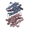
| Title | R2F from Corynebacterium Ammoniagenes in its reduced, Fe containing, form | ||||||
 Components Components | Ribonucleotide reductase protein R2F | ||||||
 Keywords Keywords | METAL BINDING PROTEIN / helix bundle / arm exchange / radical protein | ||||||
| Function / homology |  Function and homology information Function and homology informationribonucleoside-diphosphate reductase complex / ribonucleoside-diphosphate reductase / ribonucleoside-diphosphate reductase activity, thioredoxin disulfide as acceptor / deoxyribonucleotide biosynthetic process / metal ion binding Similarity search - Function | ||||||
| Biological species |  Corynebacterium ammoniagenes (bacteria) Corynebacterium ammoniagenes (bacteria) | ||||||
| Method |  X-RAY DIFFRACTION / SYNCHROTRON / MOLECULAR REPLACEMENT / Resolution: 2.25 Å X-RAY DIFFRACTION / SYNCHROTRON / MOLECULAR REPLACEMENT / Resolution: 2.25 Å | ||||||
 Authors Authors | Hogbom, M. / Huque, Y. / Sjoberg, B.M. / Nordlund, P. | ||||||
 Citation Citation | Journal: Biochemistry / Year: 2002 Title: Crystal structure of the di-iron/radical protein of ribonucleotide reductase from Corynebacterium ammoniagenes. Authors: Hogbom, M. / Huque, Y. / Sjoberg, B.M. / Nordlund, P. | ||||||
| History |
|
- Structure visualization
| Structure viewer | Molecule: MolmilJmol/JSmol |
|---|
- Downloads & links
-Download
| PDBx/mmCIF format | 1kgo.cif.gz | 266 KB | Display | PDBx/mmCIF format |
|---|---|---|---|---|
| PDB format | pdb1kgo.ent.gz | 214.6 KB | Display | PDB format |
| PDBx/mmJSON format | 1kgo.json.gz | Tree view | PDBx/mmJSON format | |
| Others |  Other downloads Other downloads |
-Validation report
| Arichive directory | https://data.pdbj.org/pub/pdb/validation_reports/kg/1kgoftp://data.pdbj.org/pub/pdb/validation_reports/kg/1kgo | HTTPS FTP |
|---|
-Related structure data


-Links
 PDBj
PDBj


- Assembly
| Deposited unit | 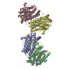
| ||||||||
|---|---|---|---|---|---|---|---|---|---|
| 1 | 
| ||||||||
| 2 | 
| ||||||||
| Unit cell |
| ||||||||
| Details | The biological assembly is one dimer. The asymmetric unit contains two of these dimers |
-Components
| #1: Protein | Mass: 37959.023 Da / Num. of mol.: 4 Source method: isolated from a genetically manipulated source Source: (gene. exp.) Corynebacterium ammoniagenes (bacteria)Plasmid: pIG056 / Production host: #2: Chemical | ChemComp-FE2 /   Mass: 55.845 Da / Num. of mol.: 8 / Source method: obtained synthetically / Formula: Fe Mass: 55.845 Da / Num. of mol.: 8 / Source method: obtained synthetically / Formula: Fe#3: Water | ChemComp-HOH / |  Mass: 18.015 Da / Num. of mol.: 848 / Source method: isolated from a natural source / Formula: H2O Mass: 18.015 Da / Num. of mol.: 848 / Source method: isolated from a natural source / Formula: H2O |
|---|
-Experimental details
-Experiment
| Experiment | Method: X-RAY DIFFRACTION / Number of used crystals: 1 |
|---|
- Sample preparation
| Crystal | Density Matthews: 2.06 Å3/Da / Density % sol: 40.21 % | ||||||||||||||||||||||||||||||||||||
|---|---|---|---|---|---|---|---|---|---|---|---|---|---|---|---|---|---|---|---|---|---|---|---|---|---|---|---|---|---|---|---|---|---|---|---|---|---|
| Crystal grow | Temperature: 298 K / Method: vapor diffusion, hanging drop / pH: 6.5 Details: PEG4000, sodium citrate, ammonium acetate, pH 6.5, VAPOR DIFFUSION, HANGING DROP, temperature 298K | ||||||||||||||||||||||||||||||||||||
| Crystal grow | *PLUS pH: 7.5 | ||||||||||||||||||||||||||||||||||||
| Components of the solutions | *PLUS
|
-Data collection
| Diffraction | Mean temperature: 100 K |
|---|---|
| Diffraction source | Source: SYNCHROTRON / Site: ESRF  / Beamline: ID14-2 / Wavelength: 0.933 Å / Beamline: ID14-2 / Wavelength: 0.933 Å |
| Detector | Type: ADSC QUANTUM 4 / Detector: CCD / Date: Apr 29, 2000 |
| Radiation | Monochromator: diamond (111) / Protocol: SINGLE WAVELENGTH / Monochromatic (M) / Laue (L): M / Scattering type: x-ray |
| Radiation wavelength | Wavelength: 0.933 Å / Relative weight: 1 |
| Reflection | Resolution: 2.25→25 Å / Num. all: 55206 / Num. obs: 55206 / % possible obs: 94.7 % / Observed criterion σ(F): 0 / Observed criterion σ(I): 0 / Rmerge(I) obs: 0.079 |
| Reflection shell | Resolution: 2.25→2.29 Å / Rmerge(I) obs: 0.294 / % possible all: 96.4 |
| Reflection | *PLUS Lowest resolution: 25 Å / Num. measured all: 135221 |
| Reflection shell | *PLUS % possible obs: 96.4 % |
- Processing
| Software |
| |||||||||||||||||||||||||
|---|---|---|---|---|---|---|---|---|---|---|---|---|---|---|---|---|---|---|---|---|---|---|---|---|---|---|
| Refinement | Method to determine structure: MOLECULAR REPLACEMENT / Resolution: 2.25→25 Å / σ(F): 0 / Stereochemistry target values: Engh & Huber
| |||||||||||||||||||||||||
| Refinement step | Cycle: LAST / Resolution: 2.25→25 Å
| |||||||||||||||||||||||||
| Refine LS restraints |
| |||||||||||||||||||||||||
| Xplor file |
| |||||||||||||||||||||||||
| Software | *PLUS Name: CNS / Classification: refinement | |||||||||||||||||||||||||
| Refinement | *PLUS Lowest resolution: 25 Å / σ(F): 0 / % reflection Rfree: 5 % / Rfactor obs: 0.163 | |||||||||||||||||||||||||
| Solvent computation | *PLUS | |||||||||||||||||||||||||
| Displacement parameters | *PLUS | |||||||||||||||||||||||||
| Refine LS restraints | *PLUS Type: c_bond_d / Dev ideal: 0.01 |
