Mass: 18.015 Da / Num. of mol.: 1856 / Source method: isolated from a natural source / Formula: H2O
-
Experimental details
-
Experiment
Experiment
Method: X-RAY DIFFRACTION / Number of used crystals: 1
-
Sample preparation
Crystal
Density Matthews: 2.16 Å3/Da / Density % sol: 42.52 %
Crystal grow
pH: 7 Details: 0.15-0.35 M NABR, 6-12% W/V PEG 3350 AND 0.1 M BIS TRIS PROPANE PH7 OR 0.08-0.2 M KSCN 8-15% W/V PEG 3350 AND 0.1 M BIS TRIS PROPANE, pH 7.00
Monochromator: SI(111) / Protocol: SINGLE WAVELENGTH / Monochromatic (M) / Laue (L): M / Scattering type: x-ray
Radiation wavelength
Wavelength: 0.9793 Å / Relative weight: 1
Reflection
Resolution: 1.7→39.56 Å / Num. obs: 2 / % possible obs: 100 % / Observed criterion σ(I): 1 / Redundancy: 4.1 % / Rmerge(I) obs: 0.06 / Net I/σ(I): 15.3
Reflection shell
Resolution: 1.7→1.79 Å / Redundancy: 4.1 % / Rmerge(I) obs: 0.27 / Mean I/σ(I) obs: 5.3 / % possible all: 100
-
Processing
Software
Name
Version
Classification
MOSFLM
datareduction
SCALA
datascaling
SHELXD
phasing
MLPHARE
phasing
REFMAC
5.3.0021
refinement
Refinement
Method to determine structure: SAD / Resolution: 1.7→39.56 Å / Cor.coef. Fo:Fc: 0.966 / Cor.coef. Fo:Fc free: 0.952 / SU B: 1.628 / SU ML: 0.055 / Cross valid method: THROUGHOUT / ESU R: 0.093 / ESU R Free: 0.092 / Stereochemistry target values: MAXIMUM LIKELIHOOD / Details: HYDROGENS HAVE BEEN ADDED IN THE RIDING POSITIONS.
Rfactor
Num. reflection
% reflection
Selection details
Rfree
0.188
10324
5 %
RANDOM
Rwork
0.155
-
-
-
obs
0.157
197589
100 %
-
Solvent computation
Ion probe radii: 0.8 Å / Shrinkage radii: 0.8 Å / VDW probe radii: 1.4 Å / Solvent model: MASK
 Movie
Movie Controller

 Yorodumi
 Open data
 Basic information
 Components
 Keywords
 Function and homology information
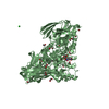
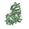
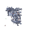
 BACTEROIDES THETAIOTAOMICRON (bacteria)
 X-RAY DIFFRACTION /
  Authors
 Citation
 Structure visualization
 Downloads & links
 Other downloads
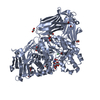
 PDBj
PDBj

 Assembly

 Mass: 282.334 Da / Num. of mol.: 3 / Source method: obtained synthetically / Formula: C11H26N2O6 / Comment: pH buffer*YM
 Mass: 92.094 Da / Num. of mol.: 9 / Source method: obtained synthetically / Formula: C3H8O3
 Mass: 35.453 Da / Num. of mol.: 4 / Source method: obtained synthetically / Formula: Cl
 Mass: 18.015 Da / Num. of mol.: 1856 / Source method: isolated from a natural source / Formula: H2O
 Sample preparation
 / Beamline: ID14-4 / Wavelength: 0.9793
  Processing