 Movie
Movie Controller

[English] 日本語
 Yorodumi
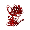
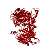
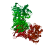
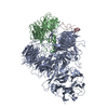
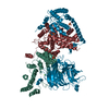
- PDB-4zli: Cellobionic acid phosphorylase - 3-O-beta-D-glucopyranosyl-alpha-... -
+ Open data

- Basic information
| Entry | Database: PDB / ID: 4zli | |||||||||
|---|---|---|---|---|---|---|---|---|---|---|
| Title | Cellobionic acid phosphorylase - 3-O-beta-D-glucopyranosyl-alpha-D-glucopyranuronic acid complex | |||||||||
 Components Components | Putative b-glycan phosphorylase | |||||||||
 Keywords Keywords | TRANSFERASE / (alpha/alpha)6 barrel / glycoside hydrolase family 94 / oxidative cellulose degradation system / intracellular enzyme | |||||||||
| Function / homology |  Function and homology information Function and homology informationhexosyltransferase activity / cellulose catabolic process / carbohydrate binding Similarity search - Function | |||||||||
| Biological species |  Saccharophagus degradans 2-40 (bacteria) Saccharophagus degradans 2-40 (bacteria) | |||||||||
| Method |  X-RAY DIFFRACTION / SYNCHROTRON / SAD / Resolution: 1.8 Å X-RAY DIFFRACTION / SYNCHROTRON / SAD / Resolution: 1.8 Å | |||||||||
 Authors Authors | Nam, Y.W. / Arakawa, T. / Fushinobu, S. | |||||||||
| Funding support |  Japan, 1items Japan, 1items
| |||||||||
 Citation Citation | Journal: J.Biol.Chem. / Year: 2015 Title: Crystal Structure and Substrate Recognition of Cellobionic Acid Phosphorylase, Which Plays a Key Role in Oxidative Cellulose Degradation by Microbes. Authors: Nam, Y.W. / Nihira, T. / Arakawa, T. / Saito, Y. / Kitaoka, M. / Nakai, H. / Fushinobu, S. #1: Journal: FEBS LETT. / Year: 2013 Title: Discovery of cellobionic acid phosphorylase in cellulolytic bacteria and fungi Authors: Nihira, T. / Saito, Y. / Nishimoto, M. / Kitaoka, M. / Igarashi, K. / Ohtsubo, K. / Nakai, H. | |||||||||
| History |
|
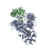
- Structure visualization
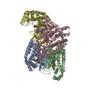
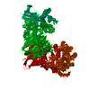
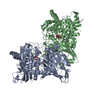
| Structure viewer | Molecule: MolmilJmol/JSmol |
|---|
- Downloads & links
-Download
| PDBx/mmCIF format | 4zli.cif.gz | 186.9 KB | Display | PDBx/mmCIF format |
|---|---|---|---|---|
| PDB format | pdb4zli.ent.gz | 144.4 KB | Display | PDB format |
| PDBx/mmJSON format | 4zli.json.gz | Tree view | PDBx/mmJSON format | |
| Others |  Other downloads Other downloads |
-Validation report
| Arichive directory | https://data.pdbj.org/pub/pdb/validation_reports/zl/4zliftp://data.pdbj.org/pub/pdb/validation_reports/zl/4zli | HTTPS FTP |
|---|
-Related structure data


-Links
 PDBj
PDBj


- Assembly
| Deposited unit | 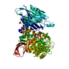
| ||||||||
|---|---|---|---|---|---|---|---|---|---|
| 1 | 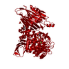
| ||||||||
| Unit cell |
| ||||||||
| Symmetry | Point symmetry: (Schoenflies symbol: C2 (2 fold cyclic)) | ||||||||
| Components on special symmetry positions |
|
-Components
-Protein / Sugars , 2 types, 2 molecules A
| #1: Protein | Mass: 89782.922 Da / Num. of mol.: 1 / Mutation: M1G Source method: isolated from a genetically manipulated source Source: (gene. exp.) Saccharophagus degradans 2-40 (bacteria)Strain: 2-40 / Gene: cep94B / Plasmid: pET28a / Production host: |
|---|---|
| #2: Polysaccharide | beta-D-glucopyranose-(1-3)-alpha-D-glucopyranuronic acid Source method: isolated from a genetically manipulated source |
-Non-polymers , 4 types, 639 molecules 


| #3: Chemical | ChemComp-GOL /  Mass: 92.094 Da / Num. of mol.: 5 / Source method: obtained synthetically / Formula: C3H8O3 Mass: 92.094 Da / Num. of mol.: 5 / Source method: obtained synthetically / Formula: C3H8O3#4: Chemical | ChemComp-SO4 /  Mass: 96.063 Da / Num. of mol.: 4 / Source method: obtained synthetically / Formula: SO4 Mass: 96.063 Da / Num. of mol.: 4 / Source method: obtained synthetically / Formula: SO4#5: Chemical | ChemComp-CL / |  Mass: 35.453 Da / Num. of mol.: 1 / Source method: obtained synthetically / Formula: Cl Mass: 35.453 Da / Num. of mol.: 1 / Source method: obtained synthetically / Formula: Cl#6: Water | ChemComp-HOH / | Mass: 18.015 Da / Num. of mol.: 629 / Source method: isolated from a natural source / Formula: H2O |
|---|
-Experimental details
-Experiment
| Experiment | Method: X-RAY DIFFRACTION |
|---|
- Sample preparation
| Crystal | Density Matthews: 3.47 Å3/Da / Density % sol: 64.5 % |
|---|---|
| Crystal grow | Temperature: 298 K / Method: vapor diffusion, sitting drop Details: 0.1 M sodium citrate (pH 5.6-6.0), 0.1 M Li2SO4, 0.6 M (NH3)2SO4, and 5% (v/v) glycerol PH range: 5.6-6.0 |
-Data collection
| Diffraction | Mean temperature: 100 K |
|---|---|
| Diffraction source | Source: SYNCHROTRON / Site: Photon Factory / Beamline: BL-17A / Wavelength: 0.9732 Å |
| Detector | Type: ADSC QUANTUM 270 / Detector: CCD / Date: Nov 29, 2013 |
| Radiation | Monochromator: Numerical link type Si(111) double crystal monochromator Protocol: SINGLE WAVELENGTH / Monochromatic (M) / Laue (L): M / Scattering type: x-ray |
| Radiation wavelength | Wavelength: 0.9732 Å / Relative weight: 1 |
| Reflection | Resolution: 1.8→50 Å / Num. obs: 114626 / % possible obs: 99.5 % / Redundancy: 7.3 % / Rsym value: 0.092 / Net I/σ(I): 24.6 |
| Reflection shell | Resolution: 1.8→1.83 Å / Redundancy: 7.4 % / Rmerge(I) obs: 0.549 / Mean I/σ(I) obs: 4.1 / % possible all: 100 |
- Processing
| Software |
| ||||||||||||||||||||||||||||||||||||||||||||||||||||||||||||||||||||||||||||||||||||||||||||||||||||||||||||||||||||||||||||||||||||||||||||||||||||||||||||||||||||||||||||||||||||||
|---|---|---|---|---|---|---|---|---|---|---|---|---|---|---|---|---|---|---|---|---|---|---|---|---|---|---|---|---|---|---|---|---|---|---|---|---|---|---|---|---|---|---|---|---|---|---|---|---|---|---|---|---|---|---|---|---|---|---|---|---|---|---|---|---|---|---|---|---|---|---|---|---|---|---|---|---|---|---|---|---|---|---|---|---|---|---|---|---|---|---|---|---|---|---|---|---|---|---|---|---|---|---|---|---|---|---|---|---|---|---|---|---|---|---|---|---|---|---|---|---|---|---|---|---|---|---|---|---|---|---|---|---|---|---|---|---|---|---|---|---|---|---|---|---|---|---|---|---|---|---|---|---|---|---|---|---|---|---|---|---|---|---|---|---|---|---|---|---|---|---|---|---|---|---|---|---|---|---|---|---|---|---|---|
| Refinement | Method to determine structure: SAD / Resolution: 1.8→40.5 Å / Cor.coef. Fo:Fc: 0.971 / Cor.coef. Fo:Fc free: 0.957 / SU B: 1.895 / SU ML: 0.058 / Cross valid method: THROUGHOUT / ESU R: 0.083 / ESU R Free: 0.087 / Stereochemistry target values: MAXIMUM LIKELIHOOD / Details: HYDROGENS HAVE BEEN ADDED IN THE RIDING POSITIONS
| ||||||||||||||||||||||||||||||||||||||||||||||||||||||||||||||||||||||||||||||||||||||||||||||||||||||||||||||||||||||||||||||||||||||||||||||||||||||||||||||||||||||||||||||||||||||
| Solvent computation | Ion probe radii: 0.8 Å / Shrinkage radii: 0.8 Å / VDW probe radii: 1.2 Å / Solvent model: BABINET MODEL WITH MASK | ||||||||||||||||||||||||||||||||||||||||||||||||||||||||||||||||||||||||||||||||||||||||||||||||||||||||||||||||||||||||||||||||||||||||||||||||||||||||||||||||||||||||||||||||||||||
| Displacement parameters | Biso mean: 28.168 Å2
| ||||||||||||||||||||||||||||||||||||||||||||||||||||||||||||||||||||||||||||||||||||||||||||||||||||||||||||||||||||||||||||||||||||||||||||||||||||||||||||||||||||||||||||||||||||||
| Refinement step | Cycle: 1 / Resolution: 1.8→40.5 Å
| ||||||||||||||||||||||||||||||||||||||||||||||||||||||||||||||||||||||||||||||||||||||||||||||||||||||||||||||||||||||||||||||||||||||||||||||||||||||||||||||||||||||||||||||||||||||
| Refine LS restraints |
|