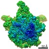
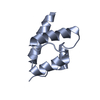
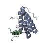
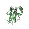
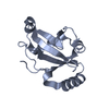
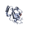
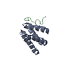
Journal: Mol Cell / Year: 2020 Title: Structures of B. subtilis Maturation RNases Captured on 50S Ribosome with Pre-rRNAs. Authors: Stephanie Oerum / Tom Dendooven / Marjorie Catala / Laetitia Gilet / Clément Dégut / Aude Trinquier / Maxime Bourguet / Pierre Barraud / Sarah Cianferani / Ben F Luisi / Ciarán Condon / Carine Tisné / Abstract: The pathways for ribosomal RNA (rRNA) maturation diverge greatly among the domains of life. In the Gram-positive model bacterium, Bacillus subtilis, the final maturation steps of the two large ...The pathways for ribosomal RNA (rRNA) maturation diverge greatly among the domains of life. In the Gram-positive model bacterium, Bacillus subtilis, the final maturation steps of the two large ribosomal subunit (50S) rRNAs, 23S and 5S pre-rRNAs, are catalyzed by the double-strand specific ribonucleases (RNases) Mini-RNase III and RNase M5, respectively. Here we present a protocol that allowed us to solve the 3.0 and 3.1 Å resolution cryoelectron microscopy structures of these RNases poised to cleave their pre-rRNA substrates within the B. subtilis 50S particle. These data provide the first structural insights into rRNA maturation in bacteria by revealing how these RNases recognize and process double-stranded pre-rRNA. Our structures further uncover how specific ribosomal proteins act as chaperones to correctly fold the pre-rRNA substrates and, for Mini-III, anchor the RNase to the ribosome. These r-proteins thereby serve a quality-control function in the process from accurate ribosome assembly to rRNA processing.
In the structure databanks used in Yorodumi, some data are registered as the other names, "COVID-19 virus" and "2019-nCoV". Here are the details of the virus and the list of structure data.
Jan 31, 2019. EMDB accession codes are about to change! (news from PDBe EMDB page)
EMDB accession codes are about to change! (news from PDBe EMDB page)
The allocation of 4 digits for EMDB accession codes will soon come to an end. Whilst these codes will remain in use, new EMDB accession codes will include an additional digit and will expand incrementally as the available range of codes is exhausted. The current 4-digit format prefixed with “EMD-” (i.e. EMD-XXXX) will advance to a 5-digit format (i.e. EMD-XXXXX), and so on. It is currently estimated that the 4-digit codes will be depleted around Spring 2019, at which point the 5-digit format will come into force.
The EM Navigator/Yorodumi systems omit the EMD- prefix.
Related info.:Q: What is EMD? / ID/Accession-code notation in Yorodumi/EM Navigator
Yorodumi is a browser for structure data from EMDB, PDB, SASBDB, etc.
This page is also the successor to EM Navigator detail page, and also detail information page/front-end page for Omokage search.
The word "yorodu" (or yorozu) is an old Japanese word meaning "ten thousand". "mi" (miru) is to see.
Related info.:EMDB / PDB / SASBDB / Comparison of 3 databanks / Yorodumi Search / Aug 31, 2016. New EM Navigator & Yorodumi / Yorodumi Papers / Jmol/JSmol / Function and homology information / Changes in new EM Navigator and Yorodumi
 Movie
Movie Controller


 Open data
 Basic information
 Components
 Keywords
 Function and homology information
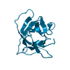
 Geobacillus stearothermophilus (bacteria)
 X-RAY DIFFRACTION /
  Authors
 France, 1items
  Citation
 Structure visualization
 Downloads & links
 Other downloads
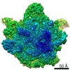


 PDBj
PDBj

 Assembly
 Mass: 18.015 Da / Num. of mol.: 98 / Source method: isolated from a natural source / Formula: H2O
 Sample preparation
 Processing