 Movie
Movie Controller


+ Open data

- Basic information
| Entry | Database: PDB / ID: 2zjd | ||||||
|---|---|---|---|---|---|---|---|
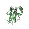
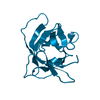
| Title | Crystal Structure of LC3-p62 complex | ||||||
 Components Components |
| ||||||
 Keywords Keywords | Apoptosis inhibitor/Apoptosis / p62 / autophagy / LC3 / microtubule-associated protein 1 light chain 3 / Cytoplasm / Cytoplasmic vesicle / Lipoprotein / Membrane / Ubl conjugation pathway / Alternative splicing / Apoptosis / Differentiation / Endosome / Immune response / Metal-binding / Nucleus / Phosphoprotein / Zinc / Zinc-finger / Apoptosis inhibitor-Apoptosis COMPLEX | ||||||
| Function / homology |  Function and homology information Function and homology informationprotein localization to perinuclear region of cytoplasm / brown fat cell proliferation / Pexophagy / NRIF signals cell death from the nucleus / p75NTR recruits signalling complexes / protein targeting to vacuole involved in autophagy / PINK1-PRKN Mediated Mitophagy / intracellular membraneless organelle / NF-kB is activated and signals survival / aggrephagy ...protein localization to perinuclear region of cytoplasm / brown fat cell proliferation / Pexophagy / NRIF signals cell death from the nucleus / p75NTR recruits signalling complexes / protein targeting to vacuole involved in autophagy / PINK1-PRKN Mediated Mitophagy / intracellular membraneless organelle / NF-kB is activated and signals survival / aggrephagy / negative regulation of toll-like receptor 4 signaling pathway / SARS-CoV-2 modulates autophagy / response to mitochondrial depolarisation / amphisome / ceramide binding / Interleukin-1 signaling / regulation of protein complex stability / endosome organization / pexophagy / phosphatidylethanolamine binding / KEAP1-NFE2L2 pathway / membraneless organelle assembly / phagophore assembly site / Translation of Replicase and Assembly of the Replication Transcription Complex / ubiquitin-modified protein reader activity / TBC/RABGAPs / regulation of canonical NF-kappaB signal transduction / aggresome / cellular response to nitrogen starvation / toll-like receptor 4 signaling pathway / K63-linked polyubiquitin modification-dependent protein binding / Receptor Mediated Mitophagy / cellular response to stress / Macroautophagy / Lewy body / organelle membrane / temperature homeostasis / negative regulation of ferroptosis / autolysosome / autophagosome membrane / molecular sequestering activity / autophagosome maturation / axoneme / protein K63-linked ubiquitination / autophagosome assembly / mitophagy / energy homeostasis / endomembrane system / inclusion body / signaling adaptor activity / negative regulation of protein ubiquitination / positive regulation of autophagy / ionotropic glutamate receptor binding / SH2 domain binding / autophagosome / response to ischemia / Pexophagy / protein kinase C binding / cellular response to starvation / protein catabolic process / positive regulation of long-term synaptic potentiation / positive regulation of protein localization to plasma membrane / PINK1-PRKN Mediated Mitophagy / sarcomere / macroautophagy / transcription coregulator activity / ubiquitin binding / protein sequestering activity / molecular condensate scaffold activity / P-body / PML body / autophagy / mitochondrial membrane / protein import into nucleus / late endosome / KEAP1-NFE2L2 pathway / signaling receptor activity / cytoplasmic vesicle / sperm midpiece / Translation of Replicase and Assembly of the Replication Transcription Complex / microtubule binding / ubiquitin-dependent protein catabolic process / microtubule / protein-macromolecule adaptor activity / cell differentiation / apoptotic process / ubiquitin protein ligase binding / synapse / protein kinase binding / protein-containing complex binding / glutamatergic synapse / enzyme binding / negative regulation of transcription by RNA polymerase II / endoplasmic reticulum / mitochondrion / zinc ion binding / identical protein binding / cytoplasm / cytosol Similarity search - Function | ||||||
| Biological species |  Homo sapiens (human) Homo sapiens (human) | ||||||
| Method |  X-RAY DIFFRACTION / SYNCHROTRON / MOLECULAR REPLACEMENT / Resolution: 1.56 Å X-RAY DIFFRACTION / SYNCHROTRON / MOLECULAR REPLACEMENT / Resolution: 1.56 Å | ||||||
 Authors Authors | Ichimura, Y. / Kumanomidou, T. / Sou, Y. / Mizushima, T. / Ezaki, J. / Ueno, T. / Kominami, E. / Yamane, T. / Tanaka, K. / Komatsu, M. | ||||||
 Citation Citation | Journal: J.Biol.Chem. / Year: 2008 Title: Structural Basis for Sorting Mechanism of p62 in Selective Autophagy Authors: Ichimura, Y. / Kumanomidou, T. / Sou, Y.-S. / Mizushima, T. / Ezaki, J. / Ueno, T. / Kominami, E. / Yamane, T. / Tanaka, K. / Komatsu, M. | ||||||
| History |
|
- Structure visualization
| Structure viewer | Molecule: MolmilJmol/JSmol |
|---|
- Downloads & links
-Download
| PDBx/mmCIF format | 2zjd.cif.gz | 71.5 KB | Display | PDBx/mmCIF format |
|---|---|---|---|---|
| PDB format | pdb2zjd.ent.gz | 53.7 KB | Display | PDB format |
| PDBx/mmJSON format | 2zjd.json.gz | Tree view | PDBx/mmJSON format | |
| Others |  Other downloads Other downloads |
-Validation report
| Arichive directory | https://data.pdbj.org/pub/pdb/validation_reports/zj/2zjdftp://data.pdbj.org/pub/pdb/validation_reports/zj/2zjd | HTTPS FTP |
|---|
-Related structure data
| Related structure data | 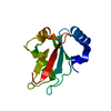 1ugmS S: Starting model for refinement |
|---|---|
| Similar structure data |


-Links
 PDBj
PDBj


- Assembly
| Deposited unit | 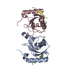
| ||||||||
|---|---|---|---|---|---|---|---|---|---|
| 1 | 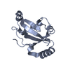
| ||||||||
| 2 | 
| ||||||||
| 3 | 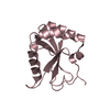
| ||||||||
| 4 | 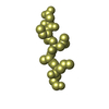
| ||||||||
| 5 | 
| ||||||||
| 6 | 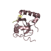
| ||||||||
| Unit cell |
|
-Components
| #1: Protein | Mass: 15121.482 Da / Num. of mol.: 2 Source method: isolated from a genetically manipulated source Source: (gene. exp.) Homo sapiens (human) / Plasmid: pGEX-6P / Production host:  #2: Protein/peptide | Mass: 1190.156 Da / Num. of mol.: 2 Fragment: LC3 recognition sequence (LRS), UNP residue 334-344 Source method: obtained synthetically / Details: This sequence occurs naturally in mouse. / References: UniProt: Q64337 #3: Water | ChemComp-HOH / |  Mass: 18.015 Da / Num. of mol.: 283 / Source method: isolated from a natural source / Formula: H2O Mass: 18.015 Da / Num. of mol.: 283 / Source method: isolated from a natural source / Formula: H2O |
|---|
-Experimental details
-Experiment
| Experiment | Method: X-RAY DIFFRACTION / Number of used crystals: 1 |
|---|
- Sample preparation
| Crystal | Density Matthews: 2.15 Å3/Da / Density % sol: 42.87 % |
|---|---|
| Crystal grow | Temperature: 288 K / Method: vapor diffusion, hanging drop / pH: 7.5 Details: 0.1M HEPES pH 7.5, 23% PEG 3350, VAPOR DIFFUSION, HANGING DROP, temperature 288K |
-Data collection
| Diffraction | Mean temperature: 100 K |
|---|---|
| Diffraction source | Source: SYNCHROTRON / Site: SPring-8  / Beamline: BL44XU / Wavelength: 0.9 Å / Beamline: BL44XU / Wavelength: 0.9 Å |
| Detector | Type: Bruker DIP-6040 / Detector: CCD / Date: Nov 21, 2007 |
| Radiation | Protocol: SINGLE WAVELENGTH / Monochromatic (M) / Laue (L): M / Scattering type: x-ray |
| Radiation wavelength | Wavelength: 0.9 Å / Relative weight: 1 |
| Reflection | Resolution: 1.55→77.85 Å / Num. obs: 39943 / % possible obs: 98.4 % / Redundancy: 4.5 % / Rmerge(I) obs: 0.054 / Net I/σ(I): 23.9 |
| Reflection shell | Resolution: 1.55→1.61 Å / Redundancy: 4.2 % / Rmerge(I) obs: 0.31 / % possible all: 86 |
- Processing
| Software |
| ||||||||||||||||||||||||||||||||||||||||||||||||||||||||||||||||||||||||||||||||||||||||||||||||||||||||||||||||||||||||||||||||||||||||||||||||||||||||||||||||||||||||||
|---|---|---|---|---|---|---|---|---|---|---|---|---|---|---|---|---|---|---|---|---|---|---|---|---|---|---|---|---|---|---|---|---|---|---|---|---|---|---|---|---|---|---|---|---|---|---|---|---|---|---|---|---|---|---|---|---|---|---|---|---|---|---|---|---|---|---|---|---|---|---|---|---|---|---|---|---|---|---|---|---|---|---|---|---|---|---|---|---|---|---|---|---|---|---|---|---|---|---|---|---|---|---|---|---|---|---|---|---|---|---|---|---|---|---|---|---|---|---|---|---|---|---|---|---|---|---|---|---|---|---|---|---|---|---|---|---|---|---|---|---|---|---|---|---|---|---|---|---|---|---|---|---|---|---|---|---|---|---|---|---|---|---|---|---|---|---|---|---|---|---|---|
| Refinement | Method to determine structure: MOLECULAR REPLACEMENT Starting model: PDB ENTRY 1UGM Resolution: 1.56→45.48 Å / Cor.coef. Fo:Fc: 0.951 / Cor.coef. Fo:Fc free: 0.931 / SU B: 1.531 / SU ML: 0.057 / Cross valid method: THROUGHOUT / ESU R: 0.093 / ESU R Free: 0.095 / Stereochemistry target values: MAXIMUM LIKELIHOOD / Details: HYDROGENS HAVE BEEN ADDED IN THE RIDING POSITIONS
| ||||||||||||||||||||||||||||||||||||||||||||||||||||||||||||||||||||||||||||||||||||||||||||||||||||||||||||||||||||||||||||||||||||||||||||||||||||||||||||||||||||||||||
| Solvent computation | Ion probe radii: 0.8 Å / Shrinkage radii: 0.8 Å / VDW probe radii: 1.2 Å / Solvent model: MASK | ||||||||||||||||||||||||||||||||||||||||||||||||||||||||||||||||||||||||||||||||||||||||||||||||||||||||||||||||||||||||||||||||||||||||||||||||||||||||||||||||||||||||||
| Displacement parameters | Biso mean: 19.107 Å2
| ||||||||||||||||||||||||||||||||||||||||||||||||||||||||||||||||||||||||||||||||||||||||||||||||||||||||||||||||||||||||||||||||||||||||||||||||||||||||||||||||||||||||||
| Refinement step | Cycle: LAST / Resolution: 1.56→45.48 Å
| ||||||||||||||||||||||||||||||||||||||||||||||||||||||||||||||||||||||||||||||||||||||||||||||||||||||||||||||||||||||||||||||||||||||||||||||||||||||||||||||||||||||||||
| Refine LS restraints |
| ||||||||||||||||||||||||||||||||||||||||||||||||||||||||||||||||||||||||||||||||||||||||||||||||||||||||||||||||||||||||||||||||||||||||||||||||||||||||||||||||||||||||||
| LS refinement shell | Resolution: 1.556→1.596 Å / Total num. of bins used: 20
|
