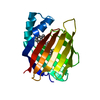
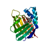
Journal: Biochemistry / Year: 1998 Title: NMR study suggests a major role for Arg111 in maintaining the structure and dynamical properties of type II human cellular retinoic acid binding protein. Authors: Wang, L. / Yan, H.
Mass: 15555.804 Da / Num. of mol.: 1 / Mutation: R111M Source method: isolated from a genetically manipulated source Source: (gene. exp.) Homo sapiens (human) / Cell line: BL21 / Plasmid: PET-17B / Cellular location (production host): CYTOPLASM / Production host: Escherichia coli (E. coli) / Strain (production host): BL21 (DE3) PLYSS / References: UniProt: P29373
-
Experimental details
-
Experiment
Experiment
Method: SOLUTION NMR
NMR experiment
Conditions-ID
Experiment-ID
Solution-ID
Type
1
1
1
2DHOMONUCLEAR
1
2
1
3D 15N-EDITED NMR EXPERIMENTS
-
Sample preparation
Details
Contents: PBS
Sample conditions
pH: 7.3 / Temperature: 300 K
Crystal grow
*PLUS
Method: other / Details: NMR
-
NMR measurement
NMR spectrometer
Type
Manufacturer
Model
Field strength (MHz)
Spectrometer-ID
Varian VXR500
Varian
VXR500
500
1
Bruker DMX750
Bruker
DMX750
750
2
-
Processing
Software
Name
Version
Classification
X-PLOR
3.1
modelbuilding
X-PLOR
3.1
refinement
X-PLOR
3.1
phasing
NMR software
Name
Version
Developer
Classification
X-PLOR
3.1
BRUNGER
refinement
X-PLOR
structuresolution
Refinement
Method: DGSA / Software ordinal: 1 Details: THE STRUCTURES WERE CALCULATED FOLLOWING THE STANDARD DISTANCE GEOMETRY-SIMULATED ANNEALING PROTOCAL IN X-PLOR 3.1. SEVERAL ROUNDS OF REFINEMENT WERE PERFORMED.
NMR ensemble
Conformer selection criteria: LEAST RESTRAINT VIOLATION AND LOWEST TOTAL ENERGY Conformers calculated total number: 75 / Conformers submitted total number: 31
+
About Yorodumi
-
News
-
Feb 9, 2022. New format data for meta-information of EMDB entries
New format data for meta-information of EMDB entries
Version 3 of the EMDB header file is now the official format.
The previous official version 1.9 will be removed from the archive.
In the structure databanks used in Yorodumi, some data are registered as the other names, "COVID-19 virus" and "2019-nCoV". Here are the details of the virus and the list of structure data.
Jan 31, 2019. EMDB accession codes are about to change! (news from PDBe EMDB page)
EMDB accession codes are about to change! (news from PDBe EMDB page)
The allocation of 4 digits for EMDB accession codes will soon come to an end. Whilst these codes will remain in use, new EMDB accession codes will include an additional digit and will expand incrementally as the available range of codes is exhausted. The current 4-digit format prefixed with “EMD-” (i.e. EMD-XXXX) will advance to a 5-digit format (i.e. EMD-XXXXX), and so on. It is currently estimated that the 4-digit codes will be depleted around Spring 2019, at which point the 5-digit format will come into force.
The EM Navigator/Yorodumi systems omit the EMD- prefix.
Related info.:Q: What is EMD? / ID/Accession-code notation in Yorodumi/EM Navigator
Yorodumi is a browser for structure data from EMDB, PDB, SASBDB, etc.
This page is also the successor to EM Navigator detail page, and also detail information page/front-end page for Omokage search.
The word "yorodu" (or yorozu) is an old Japanese word meaning "ten thousand". "mi" (miru) is to see.
Related info.:EMDB / PDB / SASBDB / Comparison of 3 databanks / Yorodumi Search / Aug 31, 2016. New EM Navigator & Yorodumi / Yorodumi Papers / Jmol/JSmol / Function and homology information / Changes in new EM Navigator and Yorodumi
 Movie
Movie Controller

 Yorodumi
 Open data
 Basic information
 Components
 Keywords
 Function and homology information
 Homo sapiens (human)
 Authors
 Citation
 Structure visualization
 Downloads & links
 Other downloads


 PDBj
PDBj


 Assembly

 Sample preparation
 Processing
 X-PLOR