 Movie
Movie Controller

[English] 日本語
 Yorodumi
- PDB-6mea: Crystal structure of a Tylonycteris bat coronavirus HKU4 macrodom... -
+ Open data

- Basic information
| Entry | Database: PDB / ID: 6mea | ||||||
|---|---|---|---|---|---|---|---|
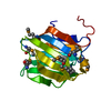
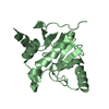
| Title | Crystal structure of a Tylonycteris bat coronavirus HKU4 macrodomain in complex with adenosine diphosphate ribose (ADP-ribose) | ||||||
 Components Components | Replicase polyprotein 1ab | ||||||
 Keywords Keywords | HYDROLASE / Coronavirus / Macrodomain / ADP-ribose / ADP-ribosylhydrolase | ||||||
| Function / homology |  Function and homology information Function and homology informationhost cell membrane / Lyases; Phosphorus-oxygen lyases / endonuclease activity / Hydrolases; Acting on ester bonds; Exoribonucleases producing 5'-phosphomonoesters / host cell endoplasmic reticulum-Golgi intermediate compartment / 3'-5'-RNA exonuclease activity / symbiont-mediated degradation of host mRNA / 5'-3' DNA helicase activity / mRNA guanylyltransferase / symbiont-mediated suppression of host ISG15-protein conjugation ...host cell membrane / Lyases; Phosphorus-oxygen lyases / endonuclease activity / Hydrolases; Acting on ester bonds; Exoribonucleases producing 5'-phosphomonoesters / host cell endoplasmic reticulum-Golgi intermediate compartment / 3'-5'-RNA exonuclease activity / symbiont-mediated degradation of host mRNA / 5'-3' DNA helicase activity / mRNA guanylyltransferase / symbiont-mediated suppression of host ISG15-protein conjugation / G-quadruplex RNA binding / mRNA guanylyltransferase activity / omega peptidase activity / mRNA (guanine-N7)-methyltransferase / methyltransferase cap1 / DNA helicase / symbiont-mediated suppression of host NF-kappaB cascade / symbiont-mediated perturbation of host ubiquitin-like protein modification / methyltransferase cap1 activity / mRNA 5'-cap (guanine-N7-)-methyltransferase activity / cysteine-type deubiquitinase activity / ubiquitinyl hydrolase 1 / Hydrolases; Acting on peptide bonds (peptidases); Cysteine endopeptidases / RNA helicase activity / lyase activity / single-stranded RNA binding / viral protein processing / host cell perinuclear region of cytoplasm / RNA helicase / symbiont-mediated suppression of host type I interferon-mediated signaling pathway / symbiont-mediated suppression of host gene expression / viral translational frameshifting / symbiont-mediated activation of host autophagy / RNA-directed RNA polymerase / cysteine-type endopeptidase activity / viral RNA genome replication / RNA-directed RNA polymerase activity / DNA-templated transcription / ATP hydrolysis activity / proteolysis / zinc ion binding / ATP binding Similarity search - Function | ||||||
| Biological species |  Bat coronavirus HKU4 Bat coronavirus HKU4 | ||||||
| Method |  X-RAY DIFFRACTION / SYNCHROTRON / MOLECULAR REPLACEMENT / Resolution: 1.35 Å X-RAY DIFFRACTION / SYNCHROTRON / MOLECULAR REPLACEMENT / Resolution: 1.35 Å | ||||||
 Authors Authors | Hammond, R.G. / Schormann, N. / McPherson, R.L. / Leung, A.K.L. / Deivanayagam, C.C.S. / Johnson, M.A. | ||||||
| Funding support |  United States, 1items United States, 1items
| ||||||
 Citation Citation | Journal: Proc.Natl.Acad.Sci.USA / Year: 2021 Title: ADP-Ribose and Analogues bound to the DeMARylating Macrodomain from the Bat Coronavirus HKU4 Authors: Hammond, R.G. / Schormann, N. / McPherson, R.L. / Leung, A.K.L. / Deivanayagam, C. / Johnson, M.A. | ||||||
| History |
|
- Structure visualization
| Structure viewer | Molecule: MolmilJmol/JSmol |
|---|
- Downloads & links
-Download
| PDBx/mmCIF format | 6mea.cif.gz | 153.3 KB | Display | PDBx/mmCIF format |
|---|---|---|---|---|
| PDB format | pdb6mea.ent.gz | 119 KB | Display | PDB format |
| PDBx/mmJSON format | 6mea.json.gz | Tree view | PDBx/mmJSON format | |
| Others |  Other downloads Other downloads |
-Validation report
| Arichive directory | https://data.pdbj.org/pub/pdb/validation_reports/me/6meaftp://data.pdbj.org/pub/pdb/validation_reports/me/6mea | HTTPS FTP |
|---|
-Related structure data
| Related structure data |  6mebC 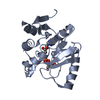 6menC  5dusS C: citing same article ( S: Starting model for refinement |
|---|---|
| Similar structure data |


-Links
 PDBj
PDBj


- Assembly
| Deposited unit | 
| ||||||||
|---|---|---|---|---|---|---|---|---|---|
| 1 | 
| ||||||||
| 2 | 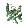
| ||||||||
| Unit cell |
|
-Components
| #1: Protein | Mass: 18595.129 Da / Num. of mol.: 2 / Fragment: macrodomain (UNP residues 1154-1324) Source method: isolated from a genetically manipulated source Source: (gene. exp.) Bat coronavirus HKU4 / Gene: rep, 1a-1b / Plasmid: pET15b-TEV / Cell line (production host): BL21(DE3)pLysS / Production host:  #2: Chemical |   Mass: 559.316 Da / Num. of mol.: 2 / Source method: obtained synthetically / Formula: C15H23N5O14P2 Mass: 559.316 Da / Num. of mol.: 2 / Source method: obtained synthetically / Formula: C15H23N5O14P2#3: Water | ChemComp-HOH / |  Mass: 18.015 Da / Num. of mol.: 372 / Source method: isolated from a natural source / Formula: H2O Mass: 18.015 Da / Num. of mol.: 372 / Source method: isolated from a natural source / Formula: H2O |
|---|
-Experimental details
-Experiment
| Experiment | Method: X-RAY DIFFRACTION / Number of used crystals: 1 |
|---|
- Sample preparation
| Crystal | Density Matthews: 2.3 Å3/Da / Density % sol: 46.46 % |
|---|---|
| Crystal grow | Temperature: 295 K / Method: vapor diffusion, hanging drop Details: 20-25% PEG3350, 0.1 M HEPES, pH 7.0-7.5, protein:ligand molar ratio 1:10 PH range: 7.0-7.5 |
-Data collection
| Diffraction | Mean temperature: 100 K |
|---|---|
| Diffraction source | Source: SYNCHROTRON / Site: APS / Beamline: 22-ID / Wavelength: 1 Å |
| Detector | Type: RAYONIX MX300-HS / Detector: CCD / Date: Jul 2, 2017 / Details: Mirrors |
| Radiation | Monochromator: double crystal Si(111) / Protocol: SINGLE WAVELENGTH / Monochromatic (M) / Laue (L): M / Scattering type: x-ray |
| Radiation wavelength | Wavelength: 1 Å / Relative weight: 1 |
| Reflection | Resolution: 1.35→39.85 Å / Num. obs: 63312 / % possible obs: 92.7 % / Redundancy: 3.7 % / Biso Wilson estimate: 10 Å2 / CC1/2: 0.991 / Rmerge(I) obs: 0.121 / Rpim(I) all: 0.074 / Rrim(I) all: 0.142 / Net I/σ(I): 9.8 |
| Reflection shell | Resolution: 1.35→1.37 Å / Redundancy: 3.4 % / Rmerge(I) obs: 0.657 / Mean I/σ(I) obs: 3 / Num. unique obs: 2865 / CC1/2: 0.662 / Rpim(I) all: 0.421 / Rrim(I) all: 0.783 / % possible all: 86.2 |
- Processing
| Software |
| |||||||||||||||||||||||||||||||||||||||||||||||||||||||||||||||||||||||||||
|---|---|---|---|---|---|---|---|---|---|---|---|---|---|---|---|---|---|---|---|---|---|---|---|---|---|---|---|---|---|---|---|---|---|---|---|---|---|---|---|---|---|---|---|---|---|---|---|---|---|---|---|---|---|---|---|---|---|---|---|---|---|---|---|---|---|---|---|---|---|---|---|---|---|---|---|---|
| Refinement | Method to determine structure: MOLECULAR REPLACEMENT Starting model: PDB entry 5DUS Resolution: 1.35→33.59 Å / Cor.coef. Fo:Fc: 0.97 / Cor.coef. Fo:Fc free: 0.965 / SU B: 1.586 / SU ML: 0.029 / Cross valid method: THROUGHOUT / σ(F): 0 / ESU R: 0.059 / ESU R Free: 0.05 Details: HYDROGENS HAVE BEEN ADDED IN THE RIDING POSITIONS U VALUES : REFINED INDIVIDUALLY
| |||||||||||||||||||||||||||||||||||||||||||||||||||||||||||||||||||||||||||
| Solvent computation | Ion probe radii: 0.8 Å / Shrinkage radii: 0.8 Å / VDW probe radii: 1.2 Å | |||||||||||||||||||||||||||||||||||||||||||||||||||||||||||||||||||||||||||
| Displacement parameters | Biso max: 52.5 Å2 / Biso mean: 14.8 Å2 / Biso min: 3.23 Å2
| |||||||||||||||||||||||||||||||||||||||||||||||||||||||||||||||||||||||||||
| Refinement step | Cycle: final / Resolution: 1.35→33.59 Å
| |||||||||||||||||||||||||||||||||||||||||||||||||||||||||||||||||||||||||||
| Refine LS restraints |
| |||||||||||||||||||||||||||||||||||||||||||||||||||||||||||||||||||||||||||
| LS refinement shell | Resolution: 1.35→1.385 Å / Rfactor Rfree error: 0 / Total num. of bins used: 20
|
