 Movie
Movie Controller


+ Open data

- Basic information
| Entry | Database: PDB / ID: 2dyc | ||||||
|---|---|---|---|---|---|---|---|
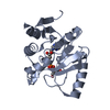
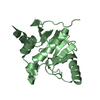
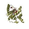
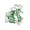
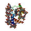
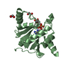
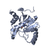
| Title | Crystal structure of the N-terminal domain of mouse galectin-4 | ||||||
 Components Components | Galectin-4 | ||||||
 Keywords Keywords | SUGAR BINDING PROTEIN / GAL-BIND LECTIN / GALECTIN / SUGAR BINDING / Structural Genomics / NPPSFA / National Project on Protein Structural and Functional Analyses / RIKEN Structural Genomics/Proteomics Initiative / RSGI | ||||||
| Function / homology |  Function and homology information Function and homology informationantibacterial peptide biosynthetic process / galactoside binding / carbohydrate binding / extracellular matrix / defense response to bacterium / : Similarity search - Function | ||||||
| Biological species |  | ||||||
| Method |  X-RAY DIFFRACTION / SYNCHROTRON / MAD / Resolution: 2.4 Å X-RAY DIFFRACTION / SYNCHROTRON / MAD / Resolution: 2.4 Å | ||||||
 Authors Authors | Kato-Murayama, M. / Murayama, K. / Terada, T. / Shirouzu, M. / Yokoyama, S. / RIKEN Structural Genomics/Proteomics Initiative (RSGI) | ||||||
 Citation Citation | Journal: To be Published Title: Crystal structure of the N-terminal domain of mouse galectin-4 Authors: Kato-Murayama, M. / Murayama, K. / Terada, T. / Shirouzu, M. / Yokoyama, S. | ||||||
| History |
|
- Structure visualization
| Structure viewer | Molecule: MolmilJmol/JSmol |
|---|
- Downloads & links
-Download
| PDBx/mmCIF format | 2dyc.cif.gz | 44.3 KB | Display | PDBx/mmCIF format |
|---|---|---|---|---|
| PDB format | pdb2dyc.ent.gz | 31.2 KB | Display | PDB format |
| PDBx/mmJSON format | 2dyc.json.gz | Tree view | PDBx/mmJSON format | |
| Others |  Other downloads Other downloads |
-Validation report
| Arichive directory | https://data.pdbj.org/pub/pdb/validation_reports/dy/2dycftp://data.pdbj.org/pub/pdb/validation_reports/dy/2dyc | HTTPS FTP |
|---|
-Related structure data
| Similar structure data | |
|---|---|
| Other databases |


-Links
 PDBj
PDBj- Assembly
| Deposited unit | 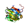
| ||||||||
|---|---|---|---|---|---|---|---|---|---|
| 1 |
| ||||||||
| 2 | x 8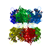
| ||||||||
| Unit cell |
|
-Components
| #1: Protein | Mass: 18149.283 Da / Num. of mol.: 1 / Fragment: residues 9-159 Source method: isolated from a genetically manipulated source Source: (gene. exp.) |
|---|---|
| #2: Water | ChemComp-HOH /  Mass: 18.015 Da / Num. of mol.: 81 / Source method: isolated from a natural source / Formula: H2O Mass: 18.015 Da / Num. of mol.: 81 / Source method: isolated from a natural source / Formula: H2O |
| Has protein modification | Y |
-Experimental details
-Experiment
| Experiment | Method: X-RAY DIFFRACTION / Number of used crystals: 1 |
|---|
- Sample preparation
| Crystal | Density Matthews: 3.88 Å3/Da / Density % sol: 68.27 % |
|---|---|
| Crystal grow | Temperature: 293 K / Method: vapor diffusion, hanging drop / pH: 7.5 Details: 1.4M Ammonium Sulfate, 12% Glycerol, Tris, pH 7.5, VAPOR DIFFUSION, HANGING DROP, temperature 293K |
-Data collection
| Diffraction | Mean temperature: 100 K | ||||||||||||
|---|---|---|---|---|---|---|---|---|---|---|---|---|---|
| Diffraction source | Source: SYNCHROTRON / Site: SPring-8  / Beamline: BL26B2 / Wavelength: 0.9790, 0.9794, 0.970 / Beamline: BL26B2 / Wavelength: 0.9790, 0.9794, 0.970 | ||||||||||||
| Detector | Type: RIGAKU JUPITER 210 / Detector: CCD / Date: Mar 7, 2006 | ||||||||||||
| Radiation | Monochromator: Si 111 CHANNEL / Protocol: MAD / Monochromatic (M) / Laue (L): M / Scattering type: x-ray | ||||||||||||
| Radiation wavelength |
| ||||||||||||
| Reflection | Resolution: 2.4→50 Å / Num. obs: 11598 / % possible obs: 99.8 % / Observed criterion σ(I): -3 / Redundancy: 9 % / Biso Wilson estimate: 21.5 Å2 / Rsym value: 0.109 / Net I/σ(I): 17.2 | ||||||||||||
| Reflection shell | Resolution: 2.4→2.49 Å / Rsym value: 0.363 / % possible all: 100 |
- Processing
| Software |
| ||||||||||||||||||||||||||||||||||||||||||||||||||||||||||||||||||||||||||||||||
|---|---|---|---|---|---|---|---|---|---|---|---|---|---|---|---|---|---|---|---|---|---|---|---|---|---|---|---|---|---|---|---|---|---|---|---|---|---|---|---|---|---|---|---|---|---|---|---|---|---|---|---|---|---|---|---|---|---|---|---|---|---|---|---|---|---|---|---|---|---|---|---|---|---|---|---|---|---|---|---|---|---|
| Refinement | Method to determine structure: MAD / Resolution: 2.4→46.5 Å / Rfactor Rfree error: 0.007 / Data cutoff high absF: 2219713.47 / Data cutoff low absF: 0 / Isotropic thermal model: RESTRAINED / Cross valid method: THROUGHOUT / σ(F): 0
| ||||||||||||||||||||||||||||||||||||||||||||||||||||||||||||||||||||||||||||||||
| Solvent computation | Solvent model: FLAT MODEL / Bsol: 36.2163 Å2 / ksol: 0.37361 e/Å3 | ||||||||||||||||||||||||||||||||||||||||||||||||||||||||||||||||||||||||||||||||
| Displacement parameters | Biso mean: 27.7 Å2
| ||||||||||||||||||||||||||||||||||||||||||||||||||||||||||||||||||||||||||||||||
| Refine analyze |
| ||||||||||||||||||||||||||||||||||||||||||||||||||||||||||||||||||||||||||||||||
| Refinement step | Cycle: LAST / Resolution: 2.4→46.5 Å
| ||||||||||||||||||||||||||||||||||||||||||||||||||||||||||||||||||||||||||||||||
| Refine LS restraints |
| ||||||||||||||||||||||||||||||||||||||||||||||||||||||||||||||||||||||||||||||||
| LS refinement shell | Resolution: 2.4→2.55 Å / Rfactor Rfree error: 0.022 / Total num. of bins used: 6
| ||||||||||||||||||||||||||||||||||||||||||||||||||||||||||||||||||||||||||||||||
| Xplor file |
|
