 Movie
Movie Controller

[English] 日本語
 Yorodumi
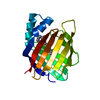
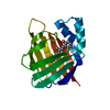
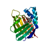
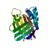
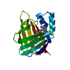
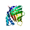
- PDB-6mqj: Crystal Structure of the All-Trans Retinal bound R111K:Y134F:T54V... -
+ Open data

- Basic information
| Entry | Database: PDB / ID: 6mqj | ||||||
|---|---|---|---|---|---|---|---|
| Title | Crystal Structure of the All-Trans Retinal bound R111K:Y134F:T54V:R132Q:P39Y:R59Y:L121Q mutant of Human Cellular Retinoic Acid Binding Protein II Irradiated with 532 nm Laser for 10 minutes at 2.1 Angstrom Resolution | ||||||
 Components Components | Cellular retinoic acid-binding protein 2 | ||||||
 Keywords Keywords | LIPID BINDING PROTEIN / iLBP / Rhodopsin mimic | ||||||
| Function / homology |  Function and homology information Function and homology informationpositive regulation of collateral sprouting / retinoid binding / retinoic acid binding / retinal binding / embryonic forelimb morphogenesis / retinoic acid metabolic process / retinol binding / Signaling by Retinoic Acid / epidermis development / fatty acid transport ...positive regulation of collateral sprouting / retinoid binding / retinoic acid binding / retinal binding / embryonic forelimb morphogenesis / retinoic acid metabolic process / retinol binding / Signaling by Retinoic Acid / epidermis development / fatty acid transport / cyclin binding / fatty acid binding / regulation of DNA-templated transcription / endoplasmic reticulum / signal transduction / extracellular exosome / nucleoplasm / nucleus / cytoplasm / cytosol Similarity search - Function | ||||||
| Biological species |  Homo sapiens (human) Homo sapiens (human) | ||||||
| Method |  X-RAY DIFFRACTION / SYNCHROTRON / MOLECULAR REPLACEMENT / Resolution: 2.125 Å X-RAY DIFFRACTION / SYNCHROTRON / MOLECULAR REPLACEMENT / Resolution: 2.125 Å | ||||||
 Authors Authors | Ghanbarpour, A. / Geiger, J. | ||||||
| Funding support |  United States, 1items United States, 1items
| ||||||
 Citation Citation | Journal: J. Am. Chem. Soc. / Year: 2019 Title: Mimicking Microbial Rhodopsin Isomerization in a Single Crystal. Authors: Ghanbarpour, A. / Nairat, M. / Nosrati, M. / Santos, E.M. / Vasileiou, C. / Dantus, M. / Borhan, B. / Geiger, J.H. | ||||||
| History |
|
- Structure visualization
| Structure viewer | Molecule: MolmilJmol/JSmol |
|---|
- Downloads & links
-Download
| PDBx/mmCIF format | 6mqj.cif.gz | 45 KB | Display | PDBx/mmCIF format |
|---|---|---|---|---|
| PDB format | pdb6mqj.ent.gz | 30.2 KB | Display | PDB format |
| PDBx/mmJSON format | 6mqj.json.gz | Tree view | PDBx/mmJSON format | |
| Others |  Other downloads Other downloads |
-Validation report
| Arichive directory | https://data.pdbj.org/pub/pdb/validation_reports/mq/6mqjftp://data.pdbj.org/pub/pdb/validation_reports/mq/6mqj | HTTPS FTP |
|---|
-Related structure data
| Related structure data |  6mopC  6moqC  6morC  6movC  6moxC  6mpkC  6mqiC  6mqwC  6mqxC  6mqyC  6mqzC  6mr0C 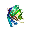 4yfpS S: Starting model for refinement C: citing same article ( |
|---|---|
| Similar structure data |


-Links
 PDBj
PDBj


- Assembly
| Deposited unit | 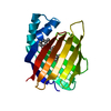
| ||||||||
|---|---|---|---|---|---|---|---|---|---|
| 1 |
| ||||||||
| 2 | 
| ||||||||
| Unit cell |
|
-Components
| #1: Protein | Mass: 15593.763 Da / Num. of mol.: 1 / Mutation: R111K,Y134F,T54V,R132Q,P39Y,R59Y,L121Q Source method: isolated from a genetically manipulated source Source: (gene. exp.) Homo sapiens (human) / Gene: CRABP2Production host: Bacterial expression vector pBEN1-SGC (others) References: UniProt: P29373 |
|---|---|
| #2: Chemical | ChemComp-RET /   Mass: 284.436 Da / Num. of mol.: 1 / Source method: obtained synthetically / Formula: C20H28O Mass: 284.436 Da / Num. of mol.: 1 / Source method: obtained synthetically / Formula: C20H28O |
| #3: Water | ChemComp-HOH /  Mass: 18.015 Da / Num. of mol.: 43 / Source method: isolated from a natural source / Formula: H2O Mass: 18.015 Da / Num. of mol.: 43 / Source method: isolated from a natural source / Formula: H2O |
| Has protein modification | Y |
-Experimental details
-Experiment
| Experiment | Method: X-RAY DIFFRACTION / Number of used crystals: 1 |
|---|
- Sample preparation
| Crystal | Density Matthews: 3.23 Å3/Da / Density % sol: 61.9 % / Description: Hexagonal |
|---|---|
| Crystal grow | Temperature: 277 K / Method: vapor diffusion, hanging drop / Details: PEG3350, DL-malic acid |
-Data collection
| Diffraction | Mean temperature: 100 K / Serial crystal experiment: N |
|---|---|
| Diffraction source | Source: SYNCHROTRON / Site: APS / Beamline: 21-ID-D / Wavelength: 0.97626 Å |
| Detector | Type: DECTRIS EIGER X 9M / Detector: PIXEL / Date: Feb 4, 2018 |
| Radiation | Protocol: SINGLE WAVELENGTH / Monochromatic (M) / Laue (L): M / Scattering type: x-ray |
| Radiation wavelength | Wavelength: 0.97626 Å / Relative weight: 1 |
| Reflection | Resolution: 2.125→45.587 Å / Num. obs: 11843 / % possible obs: 99.72 % / Redundancy: 8.7 % / Rmerge(I) obs: 0.06 / Net I/σ(I): 19.3 |
| Reflection shell | Resolution: 2.125→2.201 Å / Redundancy: 6.6 % / Rmerge(I) obs: 0.515 / Num. unique obs: 1167 / Rrim(I) all: 0.685 / % possible all: 98.9 |
- Processing
| Software |
| |||||||||||||||||||||||||||||||||||||||||||||||||||||||||||||||
|---|---|---|---|---|---|---|---|---|---|---|---|---|---|---|---|---|---|---|---|---|---|---|---|---|---|---|---|---|---|---|---|---|---|---|---|---|---|---|---|---|---|---|---|---|---|---|---|---|---|---|---|---|---|---|---|---|---|---|---|---|---|---|---|---|
| Refinement | Method to determine structure: MOLECULAR REPLACEMENT Starting model: 4YFP Resolution: 2.125→45.587 Å / SU ML: 0.26 / Cross valid method: FREE R-VALUE / σ(F): 1.34 / Phase error: 24.48 / Stereochemistry target values: ML
| |||||||||||||||||||||||||||||||||||||||||||||||||||||||||||||||
| Solvent computation | Shrinkage radii: 0.9 Å / VDW probe radii: 1.11 Å / Solvent model: FLAT BULK SOLVENT MODEL | |||||||||||||||||||||||||||||||||||||||||||||||||||||||||||||||
| Refinement step | Cycle: LAST / Resolution: 2.125→45.587 Å
| |||||||||||||||||||||||||||||||||||||||||||||||||||||||||||||||
| Refine LS restraints |
| |||||||||||||||||||||||||||||||||||||||||||||||||||||||||||||||
| LS refinement shell |
|
