 Movie
Movie Controller

[English] 日本語
 Yorodumi
- PDB-4yfp: CRYSTAL STRUCTURE OF THE R111K:Y134F:T54V:R132Q:P39Y:R59Y MUTANT ... -
+ Open data

- Basic information
| Entry | Database: PDB / ID: 4yfp | ||||||
|---|---|---|---|---|---|---|---|
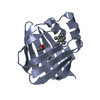
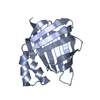
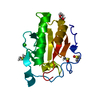
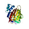
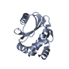
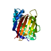
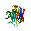
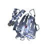
| Title | CRYSTAL STRUCTURE OF THE R111K:Y134F:T54V:R132Q:P39Y:R59Y MUTANT OF HUMAN CELLULAR RETINOIC ACID BINDING PROTEIN II WITH RETINAL AFTER 20 MINUTES INCUBATION AT 1.95 ANGSTROM RESOLUTION-Kinetic Product | ||||||
 Components Components | Cellular retinoic acid-binding protein 2 | ||||||
 Keywords Keywords | TRANSPORT PROTEIN / Photo switchable proteins / Retinal isomerization / Retinal Protonated Schiff Base pKa change / Protein engineering | ||||||
| Function / homology |  Function and homology information Function and homology informationpositive regulation of collateral sprouting / retinoid binding / retinoic acid binding / retinal binding / embryonic forelimb morphogenesis / retinoic acid metabolic process / retinol binding / Signaling by Retinoic Acid / epidermis development / fatty acid transport ...positive regulation of collateral sprouting / retinoid binding / retinoic acid binding / retinal binding / embryonic forelimb morphogenesis / retinoic acid metabolic process / retinol binding / Signaling by Retinoic Acid / epidermis development / fatty acid transport / cyclin binding / fatty acid binding / regulation of DNA-templated transcription / signal transduction / endoplasmic reticulum / extracellular exosome / nucleoplasm / nucleus / cytoplasm / cytosol Similarity search - Function | ||||||
| Biological species |  Homo sapiens (human) Homo sapiens (human) | ||||||
| Method |  X-RAY DIFFRACTION / SYNCHROTRON / MOLECULAR REPLACEMENT / Resolution: 1.951 Å X-RAY DIFFRACTION / SYNCHROTRON / MOLECULAR REPLACEMENT / Resolution: 1.951 Å | ||||||
 Authors Authors | Nosrati, M. / Geiger, J.H. | ||||||
 Citation Citation | Journal: J.Am.Chem.Soc. / Year: 2016 Title: A Photoisomerizing Rhodopsin Mimic Observed at Atomic Resolution. Authors: Nosrati, M. / Berbasova, T. / Vasileiou, C. / Borhan, B. / Geiger, J.H. | ||||||
| History |
|
- Structure visualization
| Structure viewer | Molecule: MolmilJmol/JSmol |
|---|
- Downloads & links
-Download
| PDBx/mmCIF format | 4yfp.cif.gz | 77.2 KB | Display | PDBx/mmCIF format |
|---|---|---|---|---|
| PDB format | pdb4yfp.ent.gz | 57.6 KB | Display | PDB format |
| PDBx/mmJSON format | 4yfp.json.gz | Tree view | PDBx/mmJSON format | |
| Others |  Other downloads Other downloads |
-Validation report
| Arichive directory | https://data.pdbj.org/pub/pdb/validation_reports/yf/4yfpftp://data.pdbj.org/pub/pdb/validation_reports/yf/4yfp | HTTPS FTP |
|---|
-Related structure data
| Related structure data | 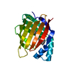 4ybpC 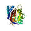 4ybuC 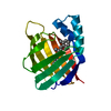 4yceC 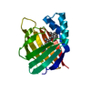 4ychC 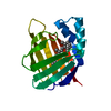 4ydaC 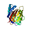 4ydbC  4yfqC 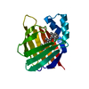 4yfrC 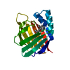 4yggC 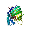 4yghC 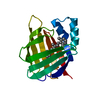 4ygzC 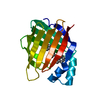 4yh0C  4ykmC  4ykoC  2g7bS C: citing same article ( S: Starting model for refinement |
|---|---|
| Similar structure data |


-Links
 PDBj
PDBj


- Assembly
| Deposited unit | 
| ||||||||
|---|---|---|---|---|---|---|---|---|---|
| 1 |
| ||||||||
| 2 | 
| ||||||||
| Unit cell |
|
-Components
| #1: Protein | Mass: 15578.792 Da / Num. of mol.: 1 / Mutation: R111K, Y134F, T54V, R132Q, P39Y, R59Y Source method: isolated from a genetically manipulated source Source: (gene. exp.) Homo sapiens (human) / Gene: CRABP2 / Plasmid: Pet17b / Production host:  |
|---|---|
| #2: Chemical | ChemComp-RET /   Mass: 284.436 Da / Num. of mol.: 1 / Source method: obtained synthetically / Formula: C20H28O Mass: 284.436 Da / Num. of mol.: 1 / Source method: obtained synthetically / Formula: C20H28O |
| #3: Water | ChemComp-HOH /  Mass: 18.015 Da / Num. of mol.: 117 / Source method: isolated from a natural source / Formula: H2O Mass: 18.015 Da / Num. of mol.: 117 / Source method: isolated from a natural source / Formula: H2O |
| Has protein modification | Y |
-Experimental details
-Experiment
| Experiment | Method: X-RAY DIFFRACTION |
|---|
- Sample preparation
| Crystal | Density Matthews: 3.22 Å3/Da / Density % sol: 61.76 % |
|---|---|
| Crystal grow | Temperature: 277 K / Method: vapor diffusion, hanging drop / pH: 6 / Details: 12% PEG3350, 0.1M Malonate pH 6.0 |
-Data collection
| Diffraction | Mean temperature: 100 K |
|---|---|
| Diffraction source | Source: SYNCHROTRON / Site: APS  / Beamline: 21-ID-F / Wavelength: 0.9787 Å / Beamline: 21-ID-F / Wavelength: 0.9787 Å |
| Detector | Type: MARMOSAIC 225 mm CCD / Detector: CCD / Date: Oct 14, 2012 |
| Radiation | Protocol: SINGLE WAVELENGTH / Monochromatic (M) / Laue (L): M / Scattering type: x-ray |
| Radiation wavelength | Wavelength: 0.9787 Å / Relative weight: 1 |
| Reflection | Resolution: 1.95→50 Å / Num. obs: 15215 / % possible obs: 99.5 % / Redundancy: 10.8 % / Rmerge(I) obs: 0.094 / Rsym value: 0.08 / Net I/σ(I): 34.6 |
| Reflection shell | Resolution: 1.95→1.98 Å / Redundancy: 10.4 % / Rmerge(I) obs: 0.639 / Mean I/σ(I) obs: 3 / % possible all: 99.4 |
- Processing
| Software |
| ||||||||||||||||||||||||||||||||||||||||||
|---|---|---|---|---|---|---|---|---|---|---|---|---|---|---|---|---|---|---|---|---|---|---|---|---|---|---|---|---|---|---|---|---|---|---|---|---|---|---|---|---|---|---|---|
| Refinement | Method to determine structure: MOLECULAR REPLACEMENT Starting model: 2G7B Resolution: 1.951→35.709 Å / SU ML: 0.5 / Cross valid method: FREE R-VALUE / σ(F): 1.38 / Phase error: 19.45 / Stereochemistry target values: ML
| ||||||||||||||||||||||||||||||||||||||||||
| Solvent computation | Shrinkage radii: 0.98 Å / VDW probe radii: 1.2 Å / Solvent model: FLAT BULK SOLVENT MODEL / Bsol: 44.719 Å2 / ksol: 0.333 e/Å3 | ||||||||||||||||||||||||||||||||||||||||||
| Displacement parameters |
| ||||||||||||||||||||||||||||||||||||||||||
| Refinement step | Cycle: LAST / Resolution: 1.951→35.709 Å
| ||||||||||||||||||||||||||||||||||||||||||
| Refine LS restraints |
| ||||||||||||||||||||||||||||||||||||||||||
| LS refinement shell |
| ||||||||||||||||||||||||||||||||||||||||||
| Refinement TLS params. | Method: refined / Origin x: -23.4834 Å / Origin y: 12.5026 Å / Origin z: -4.8679 Å
| ||||||||||||||||||||||||||||||||||||||||||
| Refinement TLS group | Selection details: all |
